Istanbul Retina Institute
FULL-THICKNESS MACULAR HOLE is an anatomic defect in the fovea featuring interruption of all neural retinal layers from the internal limiting membrane to the retina pigment epithelium.
Classification of macular hole:
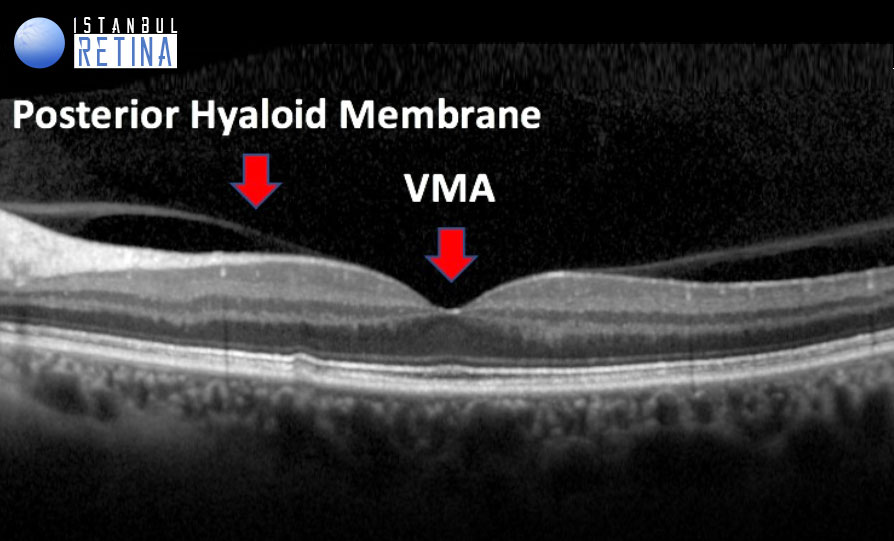
Stage 0 Macular Hole (Premacular Hole): Premacular hole can be defined as persistent foveal adhesion accompanying posterior vitreous detachment. It corresponds to vitreomacular adhesion (VMA).
Stage 1 Macular Hole: Based-on OCT evaluation, it is divided into Stage 1A and Stage 1B macular hole
STAGE 1A MACULAR HOLE: Cases with foveal pseudocyst or horizontal separation (schisis) accompanying vitreous traction in the fovea.

STAGE 1B MACULAR HOLE: Outer retinal defect accompanying vitreous traction in the fovea

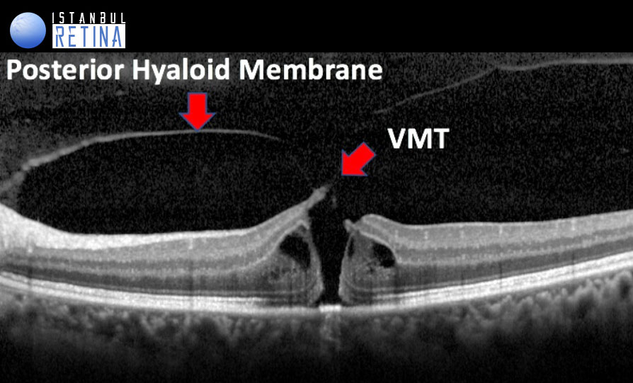
Stage 2 Macular Hole: An early full thickness macular hole with an aperture size of less than 400 µm with vitreomacular traction.
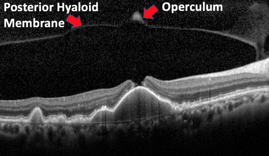
Stage 3 Macular Hole is a full-thickness macular hole larger than 400 µm in diameter and typically surrounded by a thick and detached retina at the edges. The posterior hyaloid is attached to the optic nerve head, but separated from the fovea. The operculum can be seen above the hole. In OCT, a medium or large full thickness macular hole not accompanied by VMT in the fovea is seen.


Stage 4 macular hole is a full thickness macular hole with posterior vitreous detachment and Weiss ring. In OCT, a large macular hole without vitreomacular traction is seen.

Optical Coherence TomographyeBased Full-Thickness Macular Hole Classification System (Size of Hole, Presence or Absence of Vitreomacular Traction, Cause)
In this OCT-based anatomic classification system, macular holes are determined to be small, medium, or large based on aperture size.
A small FTMH features an aperture size of less than 250 µm
A medium FTMH is defined by aperture size from 250 to 400 µm
A large FTMH is defined by aperture size >400 µm
Primary Versus Secondary Full-Thickness Macular Hole.
Full-thickness macular hole can be subdivided into primary and secondary forms. Primary FTMH (formerly referred to as idiopathic) results from vitreous traction on the fovea from anomalous PVD. A secondary FTMH is caused directly by other pathologic features and does not have pre- existing or concurrent VMT. A partial list of conditions that have been shown to result in secondary macular hole includes the following: (1) blunt trauma, (2) lightning strike, (3) high myopia, (4) macular schisis, (5) macular telangiectasia type 2, (6) wet macular degeneration treated with antievascular endothelial growth factor therapy .
Status of the Vitreous
Presence or Absence of Vitreomacular Traction. In the OCT-based anatomic system, FTMHs are categorized secondarily according to absence or presence of vitreous attachment


REFERENCES
Duker JS, Kaiser PK, Binder S, et all. The International Vitreomacular Traction Study Group Classification of Vitreomacular Adhesion, Traction, and Macular Hole. Ophthalmology 2013;120:2611-2619
Karacorlu M, Sayman Muslubas I, Ersoz MG, Hocaoglu M, Arf S.When does visual acuity stabilize after macular hole surgery? Five-year follow-up of surgery for idiopathic macular hole. Acta Ophthalmol. 2019 Feb;97(1):e136-e137
Hocaoglu M, Muslubas IS, Ersoz MG, Arf S, Karacorlu M. First-Operated and Fellow Eyes With Bilateral Idiopathic Macular Hole: Comparison of Anatomical and Functional Postoperative Outcomes. Ophthalmic Surg Lasers Imaging Retina. 2018 Aug 1;49(8):571-578
Karacorlu M, Sayman Muslubas I, Hocaoglu M, Arf S, Ersoz MG.DOUBLE ARCUATE RELAXING RETINOTOMY FOR A LARGE MACULAR HOLE. Retin Cases Brief Rep. 2019 Spring;13(2):167-170.