Istanbul Retina Institute
The most common pathology of the vitreoretinal interface is the separation of the posterior cortical vitreous from the internal limiting membrane. This is called “POSTERIOR VITREUS DETACHMENT” and this situation can be localized, partial or total.
However, in the classification scheme based on OCT, the term “VITREOMACULAR ADHESION” represents
1. A specific stage of vitreous separation
2. Partial detachment of the vitreous in the perifoveal area
3. Without retinal anomalies.
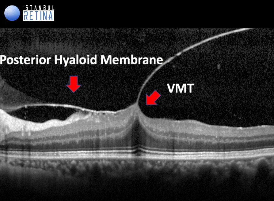

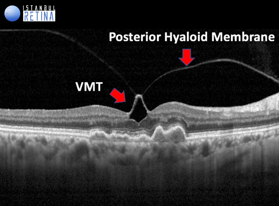
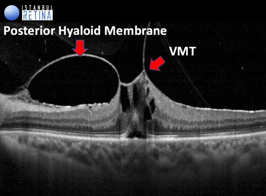
Most eyes have complete vitreoretinal adhesion at birth, so the concept of vitreoretinal adhesion and VMA is a normal state. Vitreomacular adhesion is a perifoveal vitreous detachment and is defined by anatomic features detected with OCT. Vitreomacular adhesion is characterized by an elevation of the cortical vitreous above the retinal surface, with the vitreous remaining attached within a 3-mm radius of the fovea. The angle between the vitreous and the inner retinal surface is acute, and the retina displays no change in contour or morphologic features on OCT because of the vitreous adhesion. People with VMA generally experience no visual impairment, and the finding is normal in the natural course of PVD.
Eyes with VMA may be subclassified by size of the adhesion into either: FOCAL (≤1500μm) or BROAD (>1500 mm). Eyes with VMA also may have other associated macular abnormalities, including age-related macular degeneration, retinal vein occlusion, or diabetic macular edema. In these eyes, VMA should be termed CONCURRENT, and the term ISOLATED should be reserved for cases where no ocular disease is present.
Duker JS, Kaiser PK, Binder S, et all. The International Vitreomacular Traction Study Group Classification of Vitreomacular Adhesion, Traction, and Macular Hole. Ophthalmology 2013;120:2611-2619