Istanbul Retina Institute
Advancements in spectral domain optical coherence tomography (SD-OCT) technology enables clear visualization of very small structural details of the posterior segment.
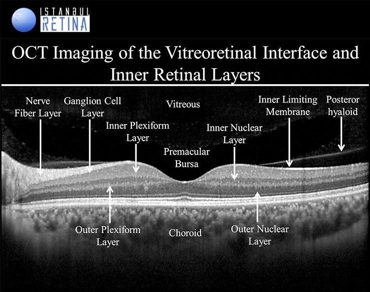
Vitreous and vitreoretinal interface:
- Vitreous (hyporeflective space)
- Premacular bursa (hyporeflective space in front of the macula)
- Posterior hyaloid (hyperreflective continuous line)
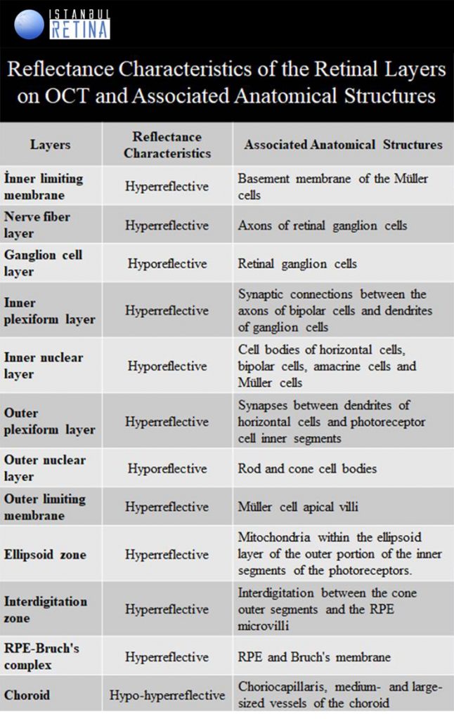
Inner retinal layers appear as hypo- and hyperreflective bands:
- Inner limiting membrane
- Nerve fiber layer
- Ganglion cell layer
- Inner plexiform layer
- Inner nuclear layer
- Outer plexiform layer
- Outer nuclear layer
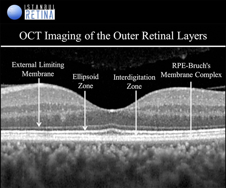
Outer retinal layers appear as four distinct hyper-reflective lines:
- External limiting membrane
- Ellipsoid zone
- Interdigitation zone
- Retinal pigment epithelium / Bruch’s membrane complex
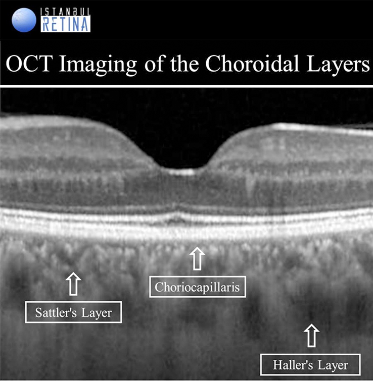
Choroid:
- Choriocapillaris (capillaries adjacent to Bruch’s membrane)
- Sattler’s layer (medium- and small-sized vessels of the choroid)
- Haller’s layer (outer choroidal layer containing large choroidal vessels)
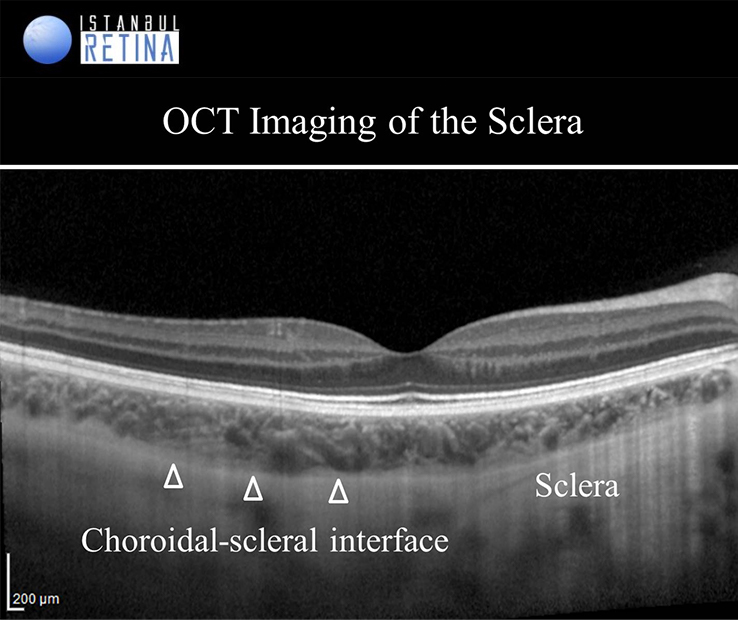
Sclera:
- Sclera is observed as a relatively uniform, hyperreflective structure exterior to the choroid on EDI-OCT images.
References:
1. Sharma P., Sergott R.C. (2016) Guide to OCT Image Interpretation with Normal and Anatomic Variants. In: Girach A., Sergott R. (eds) Optical Coherence Tomography.
2. Staurenghi G, Sadda S, Chakravarthy U, Spaide RF, International Nomenclature for Optical Coherence Tomography Panel. Proposed lexicon for anatomic landmarks in normal posterior segment spectraldomain optical coherence tomography. The IN OCT Consensus. Ophthalmology. 2014;121:1572–8.