Istanbul Retina Institute
LAMELLAR MACULAR HOLE is a partial-thickness foveal defect in which the outer retinal layers are preserved despite the defects in the inner retinal layers.
The classification of LAMELLAR MACULAR HOLE:
TRACTIONAL LAMELLAR MACULAR HOLE
Features of Tractional Lamellar Hole
- The intraretinal schisis is located between outer nuclear and outer plexiform layers
- Presence of tractional epiretinal membranes
- Intraretinal cystoid spaces in the inner plexiform layer
- Intact ellipsoid layer
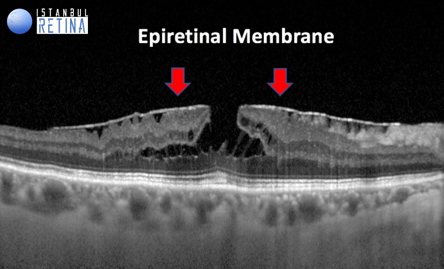
Figure 1. Tractional epiretinal membrane, intraretinal schisis, intraretinal cystic spaces and intact ellipsoid layer in a patient with lamellar macular hole.

Figure 2. Tractional epiretinal membrane, intraretinal schisis and intact ellipsoid layer in a patient with lamellar macular hole.
Degenerative Lamellar Macular Hole
Features of Degenerative Lamellar Hole
- Round-edged intraretinal cavitation
- Presence of epiretinal proliferation without traction
- No intraretinal cystic spaces
- Ellipsoid layer defect

Figure 3. Intraretinal cavitation, ellipsoid and interdigitation zone defects, epiretinal proliferation in a patient with degenerative lamellar macular hole
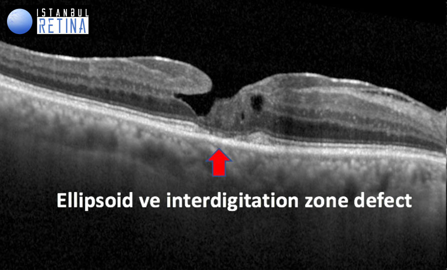
Figure 4. Ellipsoid and interdigitation zone defects and epiretinal proliferation in a patient with degenerative lamellar macular hole
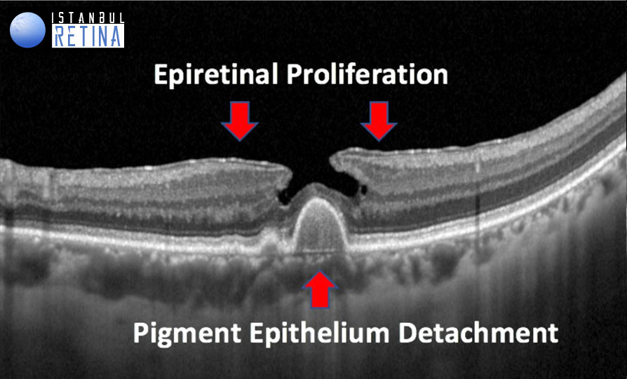
Figure 5. Intraretinal cavitation and epiretinal proliferation in a patient with degenerative lamellar macular hole and age-related macular degeneration
Macular Pseudohole
Features of Macular Pseudohole
- Invaginated or heaped foveal edges
- Concomitant ERM with central opening
- Steep macular contour to the central fovea with near-normal central foveal thickness
- No loss of retinal tissue
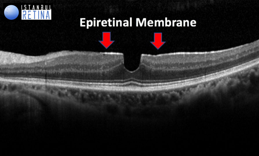
Figure 6. Epiretinal membrane in a patient with macular pseudohole

Figure 7. Epiretinal membrane in a patient with macular pseudohole
Anatomic OCT-based features of lamellar macular hole include the following: (1) an irregular foveal contour; (2) a defect in the inner fovea (may not have actual loss of tissue); (3) intraretinal splitting (schisis), typically between the outer plexiform and outer nuclear layers; and (4) features of photoreceptor layer.
In eyes with lamellar macular hole, lamellar separation of neurosensory retina demonstrated either a ‘‘cavitated’’ or a ‘‘schitic’’ appearance. The schitic appearance was defined by the presence of multiple, narrow hyperreflective tissue bridges crossing wider hyporeflective spaces, located between the outer plexiform and outer nuclear retinal layers. The cavitated appearance was defined by the presence of a homogeneous round-edged hyporeflective space in the neurosensory retina.
OCT imaging was also used to differentiate lamellar hole–associated epiretinal proliferation from classic or typical epiretinal membrane. The classical epiretinal membrane tissue was diagnosed as a thinner, irregular and hyperreflective line on the inner retinal surface, occasionally accompanied by areas of hyporeflective space between the membrane and the inner retina, while the lamellar macular hole–associated proliferation was defined as thicker preretinal material of homogenous medium reflectivity.
The presence of epiretinal proliferation and ellipsoid defect is noticeable at early stages of degenerative lamellar macular hole formation, without signs of traction. The pathophysiological process seems slow but progressive, and involves all retinal layers.
References
Duker JS, Kaiser PK, Binder S, et al. The International Vitreomacular Traction Study Group Classification of Vitreomacular Adhesion, Traction, and Macular Hole. Ophthalmology 2013;120:2611-2619
Govetto A, Dacquay Y, Farajzadeh M, et al. Lamellar Macular Hole: Two Distinct Clinical Entities? Am J Ophthalmol 2016;164:99–109
Hocaoglu M, Sayman Muslubas I, Ozdemır MH, Arf S, Karacorlu M. Lamellar Maküler Defektler. Retina Vitreus, 2015;23(1), 88-91.