Medical History:
A 31-year-old male patient with bilateral impaired vision from childhood was examined in our clinic.
Diabetes mellitus (-)
Systemic hypertension (-)
Family history (-)
Smoking (-)
Trauma (-)
Examination Findings
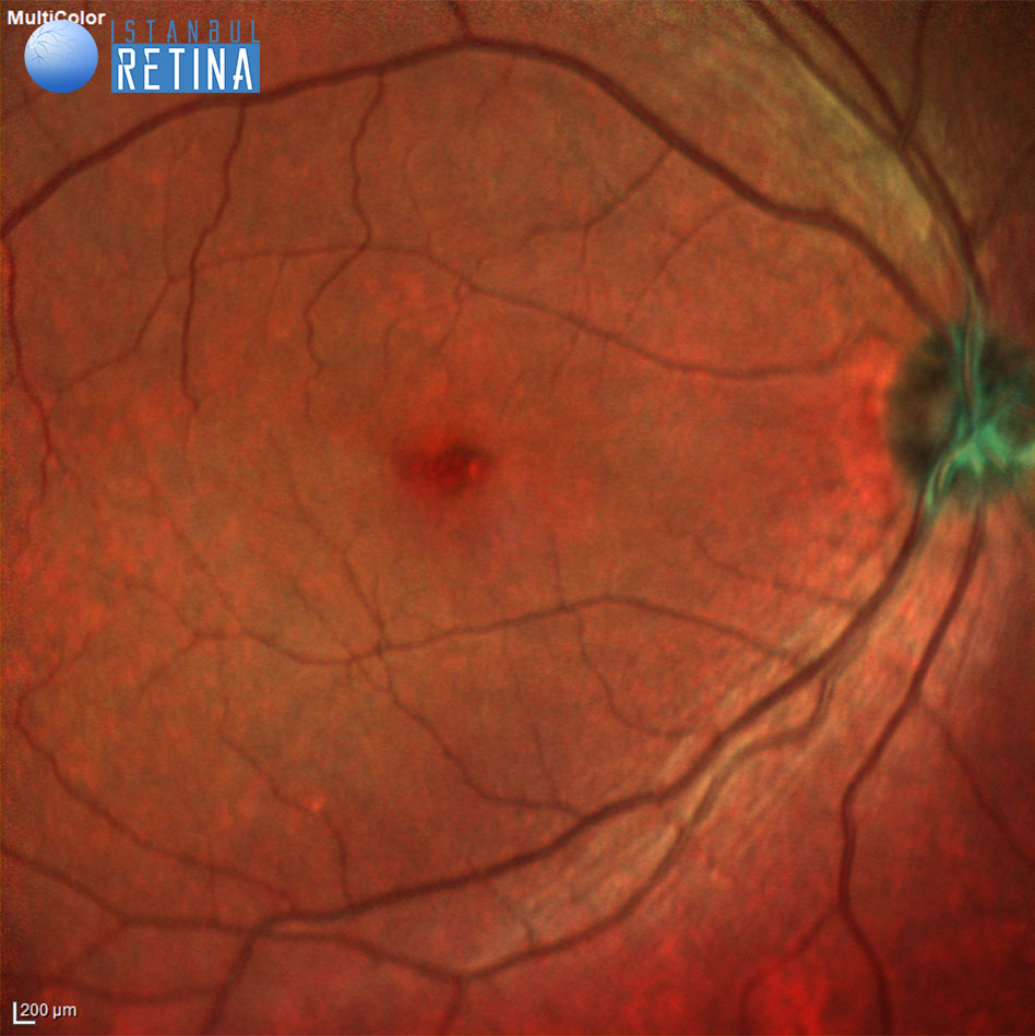
In the primary neutral position, a horizontal nystagmus was observed. The best corrected visual acuity was 1/10 in both eyes. He was not able to identify accurately the plates of Ishihara color blindness test. Intraocular pressure was 18 mmHg in both eyes. Anterior segment examination was unremarkable. Fundus examination revealed dull foveal reflex in both eyes.
Multicolor images of the patient (Figure 1).

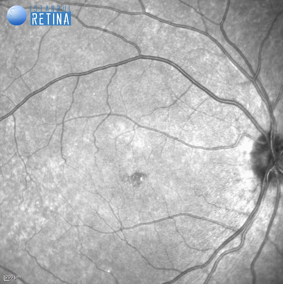
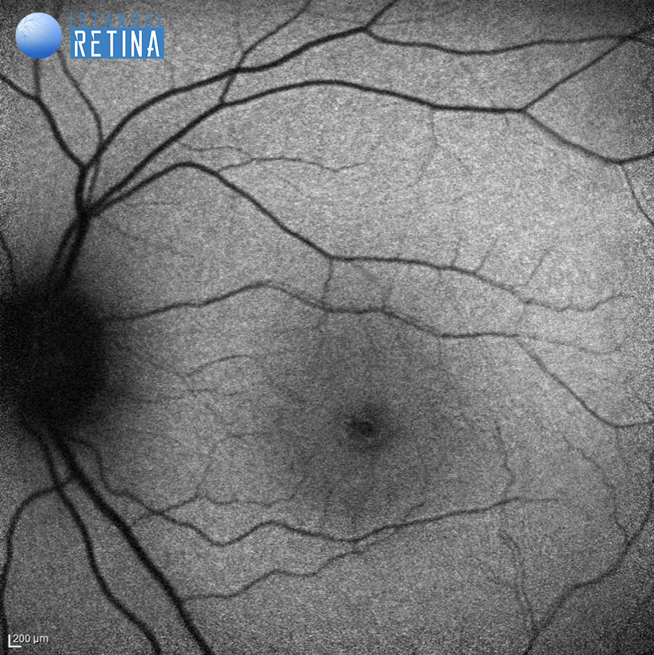
Infrared reflectance images of the patient (Figure 2).

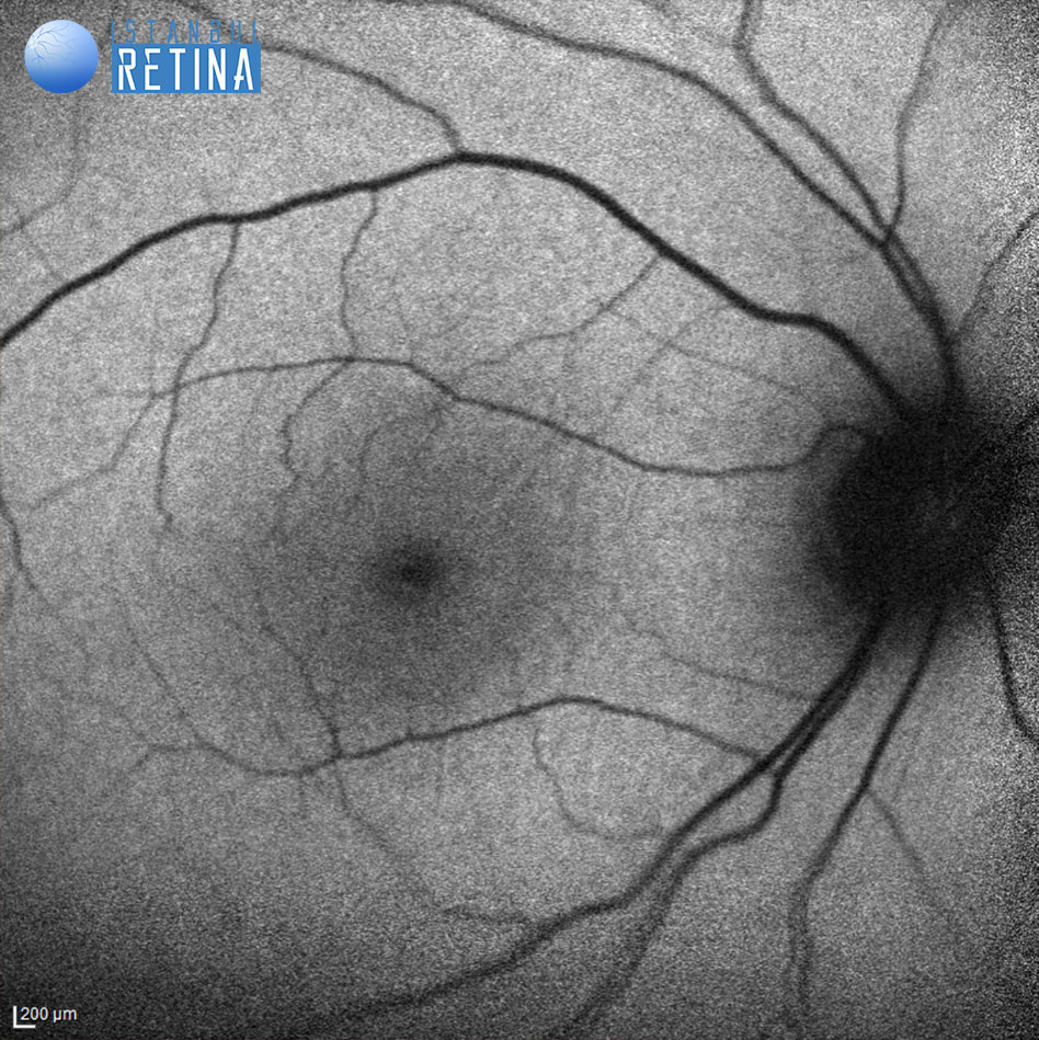
Fundus autofluorescence imaging shows reduction of normal foveal fluorescence (Figure 3).
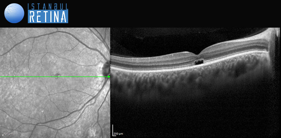
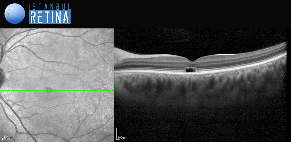
SD-OCT imaging shows presence of an optically empty space (Figure 4).
Diagnosis
Achromatopsia
Achromatopsia is the most severe form of congenital color vision deficiencies.
Complete acromatopsy with autosomal recessive inheritance is characterised by severe impairment of cone function. Autosomal recessive incomplete acromatopsy is similar to complete achromasia, however partial preservation of cone function is observed. Blue cone monochromatopsy is also known as x-linked recessive incomplete acromatopsy.
Achromatopsia was categorized into 5 stages using spectral-domain optical coherence tomography: Stage 1: intact outer retina, Stage 2: inner segment ellipsoid line disruption, Stage 3: presence of an optically empty space, Stage 4: optically empty space with partial retinal pigment epithelium disruption, Stage 5: complete retinal pigment epithelium disruption and/or loss of the outer nuclear layer.
Treatment
Currently there is no approved treatment for achromatopsia.
References
Hirji N, Aboshiha J, Georgiou M, et al. Achromatopsia: clinical features, molecular genetics, animal models and therapeutic options. Ophthalmic Genet 2018;39(2):149-157. https://pubmed.ncbi.nlm.nih.gov/29303385/
Remmer MH, Rastogi N, Ranka MP, Ceisler EJ. Achromatopsia: a review. Curr Opin Ophthalmol 2015;26(5):333-40. https://pubmed.ncbi.nlm.nih.gov/26196097/
Pascual-Camps I, Barranco-Gonzalez H, Aviñó-Martínez J, et al. Diagnosis and treatment options for achromatopsia: A Review of the Literature. J Pediatr Ophthalmol Strabismus 2018;55(2):85 92. https://pubmed.ncbi.nlm.nih.gov/29257187/
Greenberg JP, Sherman J, Zweifel SA, et al. Spectral-domain optical coherence tomography staging and autofluorescence imaging in achromatopsia. JAMA Ophthalmol 2014;132(4):437-45. https://pubmed.ncbi.nlm.nih.gov/24504161/
Aboshiha J, Dubis AM, Carroll J, et al. The cone dysfunction syndromes. Br J Ophthalmol 2016;100(1):115-21. https://pubmed.ncbi.nlm.nih.gov/25770143/