Medical History:
A 15-year-old female patient presented to our clinic complaining of decreased vision in the left eye.
Diabetes mellitus (-)
Systemic hypertension (-)
Family history (-)
Smoking (-)
Trauma (-)
Examination Findings
Best corrected visual acuity was 10/10 in the right eye and counting finger at 1 meter in the left eye. Intraocular pressure was 19 mmHg in both eyes. Anterior segment examination was normal in both eyes. Funduscopic examination revealed no pathology in the right eye and a grey-white, elevated mass located in the posterior pole in the left eye (Figure 1).
![]()
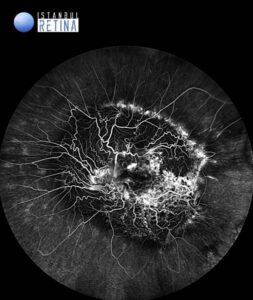
Fluorescein angiography shows hypofluorescence from blockage of choroidal vessels by RPE proliferation, abnormal vasculature with increased calibre, and diffuse late staining or leakage (Figure 2).
SD-OCT through the lesion reveals a diffusely thickened retina with intraretinal fluid and disorganisation of the inner and outer retinal architecture (Figure 3).
![]()
Diagnosis
Combined Hamartoma of the Retina and Retinal Pigment Epithelium
Combined hamartoma of retina and RPE is a rare, congenital, benign lesion which was first described by Gass in 1973. It is typically a grey-white, elevated mass located in the posterior pole involving the retina, RPE and overlying vitreous to varying degrees, frequently associated with vascular tortuosity and vitreoretinal interface changes. In the majority of patients, it is sporadic and mostly unilateral, however can be bilateral particularly when associated with neurofibromatosis type 2. Combined hamartoma of retina and RPE typically presents in childhood, most commonly with decreased visual acuity and/or strabismus. Fluorescein angiography shows early hypofluorescence from blockage of choroidal vessels by RPE proliferation, abnormal vasculature with increased calibre, arteriovenous communication, and diffuse late staining or leakage. On OCT, the lesion shows a disruption of inner neurosensory retina and variable degree of involvement of outer retina.
Differential Diagnosis
Choroidal melanoma, choroidal nevus, melanocytoma, morning glory anomaly, retinoblastoma
Treatment
The patients should be monitored for decrease in vision, ERM formation, vitreous hemorrhage and neovascularization. There are case reports of treatment of complications of combined hamartoma such as neovascularization and vitreous hemorrhage. Surgery for associated ERM is still a subject of debate since visual acuity may not improve despite membrane removal.
References:
Liu Y, Moore AT. Congenital focal abnormalities of the retina and retinal pigment epithelium. Eye (Lond). 2020;34:1973-1988. https://www.ncbi.nlm.nih.gov/pmc/articles/PMC7784997/