Medical History:
A 67-year-female patient presented to our clinic for an annual examination.
Diabetes mellitus (-)
Systemic hypertension (-)
Family history (-)
Smoking (-)
Trauma (-)
Examination Findings
Best corrected visual acuity was 10/10 in the right eye and 10/10 in the left eye. Intraocular pressure was 16 mmHg in the right eye and 15 in the left eye. Anterior segment examination was unremarkable. Fundoscopic examination of the right eye revealed hypopigmented track-like lesion temporal to the fovea (Figure 1). Fundoscopic examination of the left eye was normal.

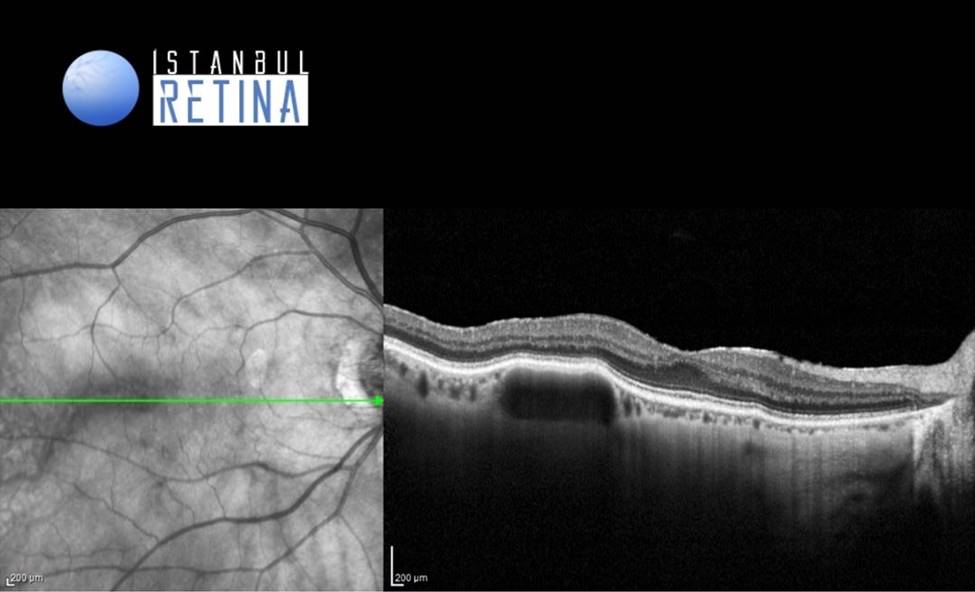
Enhanced depth imaging optical coherence tomography (OCT) revealed hyporeflective lesion occupying the entire thickness of the choroid along with the elevation of the overlying retina (Figure 2).
Diagnosis
Choroidal Macrovessel
The entity “Choroidal macrovessel” was first described in 2011 by Lima et al. Choroidal macrovessels are a rare, underdiagnosed condition mostly described in middle to older aged Caucasian women. Choroidal macrovessel is a large, tortuous anomalous vascular lesion in the choroid. In most of the cases macrovessels do not produce visual symptoms although metamorphopsia or blurry vision can occur if subretinal fluid underlying the fovea is present.
The largest case series to date by Gallo et al. demonstrated choroidal macrovessel as a hyporeflective lesion occupying the entire thickness of the choroid along with the elevation of the overlying retina, and posterior displacement of choroid-scleral junction on the OCT.
Differential Diagnosis
Ophthalmomyiasis interna, choroidal neoplasms, retinochoroidal anastomosis, and inflammatory conditions.
Treatment
Although observation is the treatment of choice, other options might be considered when subretinal fluid is present in the fovea.
References:
1. Lima LH, Laud K, Chang LK, Yannuzzi LA. Choroidal macrovessel. Br J Ophthalmol 2011;95:1333–4. https://pubmed.ncbi.nlm.nih.gov/20682950/
2. Gallo B, de Silva SR, Mahroo OA, Saihan Z, Patel PJ, Dowler JG, Pavesio C, Keane PA, Tufail A, Sagoo MS. Choroidal macrovessels: multimodal imaging findings and review of the literature. Br J Ophthalmol 2022;106:568-575. https://www.ncbi.nlm.nih.gov/pmc/articles/PMC8961769/
3. Mopuru R, Liu TYA, Arevalo JF. Choroidal Macrovessel Diagnosed on Multimodal Imaging, including Swept-Source Optical Coherence Tomography Angiography. Case Rep Ophthalmol 2022;13:215-219. https://www.ncbi.nlm.nih.gov/pmc/articles/PMC9082210/
4. Otero-Marquez O, Ledesma-Gil G, Alauddin S, Smith RT. Non-invasive imaging of a choroidal macrovessel. Am J Ophthalmol Case Rep 2020;20:100871. https://www.ncbi.nlm.nih.gov/pmc/articles/PMC7553882/