Medical History:
A 45-year-male patient presented to our clinic for an annual examination.
Diabetes mellitus (-)
Systemic hypertension (-)
Family history (-)
Smoking (-)
Trauma (-)
Examination Findings
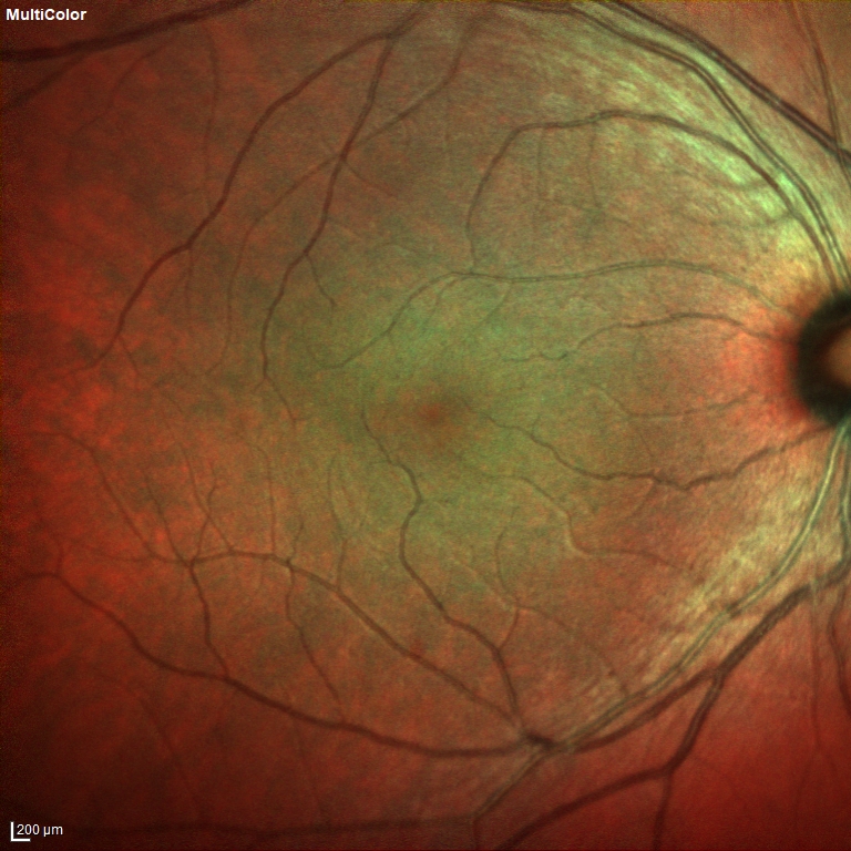
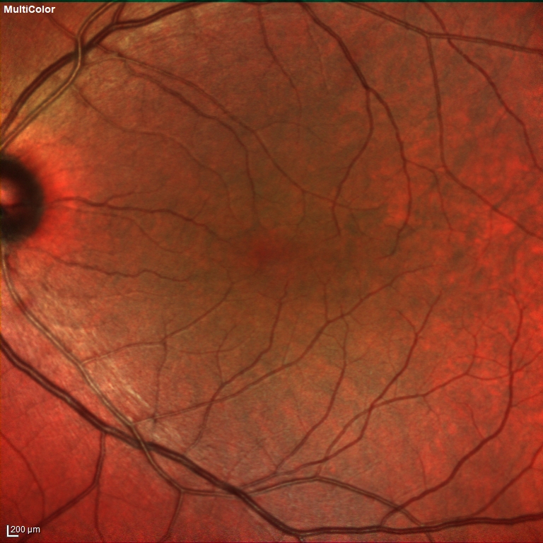
Best corrected visual acuity was 9/10 in the right eye and 9/10 in the left eye. Intraocular pressure was 13 mmHg in the right eye and 14 in the lefte ye. Anterior segment examination was unremarkable. Dilated funduscopic examination revealed normal disc and vessels (Figure 1).
The spectral domain optical coherence tomography (OCT) revealed absence of foveal pit (Figure 2).
–
–
Diagnosis
Isolated Foveal Hypoplasia
Foveal hypoplasia is a retinal disorder in which there is a lack of full development of the morphology of the fovea. It may present in isolation or be associated with other conditions such as albinism, coloboma, optic nerve hypoplasia, retinopathy of prematurity, and aniridia. Risk factors that affect foveal development, such as prematurity, can lead to foveal hypoplasia. Foveal hypoplasia has been associated with poor vision and nystagmus. An absence of a foveal pit does not necessarily imply poor visual acuity. Regardless of the degree of the development of the inner retinal layers, the visual acuity can be preserved. The mechanism causing the lack of development of the foveal pit has not been definitively determined.
Thomas et al. proposed a structural grading system of foveal hypoplasia based on the cross‑sectional OCT images: Grade 1, a shallow foveal pit and presence of outer nuclear layer and outer segment in the fovea. Grade 2, absence of foveal pit. Grade 3, absence of outer segments lengthening. Grade 4, absence of outer nuclear layer widening. Atypical form of foveal hypoplasia with disruption of the inner and outer segments of photoreceptor.
Differential Diagnosis
Albinism, aniridia, retinopathy of prematurity, incontinentia pigmenti, achromatopsia, optic nerve hypoplasia, familial exudative vitreoretinopathy, stickler syndrome
Treatment
There are no known treatments for this disorder.
References:
1.Thomas MG, Kumar A, Mohammad S, et al. Structural grading of foveal hypoplasia using spectral‑domain optical coherence tomography a predictor of visual acuity? Ophthalmology. 2011;118:1653‑60.https://www.ncbi.nlm.nih.gov/pmc/articles/PMC5648335/
2.Kondo H. Foveal hypoplasia and optical coherence tomographic imaging. Taiwan J Ophthalmol. 2018;8:181-188. https://www.ncbi.nlm.nih.gov/pmc/articles/PMC6302563/