Medical History:
A 20-year-old male patient presented with a complaint of decreased vision after blunt trauma in his left eye.
Diabetes mellitus (-)
Systemic hypertension (-)
Family history (-)
Smoking (-)
Trauma (+)
Examination Findings
Best corrected visual acuity was 10/10 in the right eye and 2/10 in the left eye. Intraocular pressure was 20 mmHg in the right eye and 25 in the left eye. Anterior segment examination was unremarkable. Fundus evaluation of the right eye was normal. Fundus examination of the left eye showed choroidal rupture and epiretinal membrane (Figure 1).

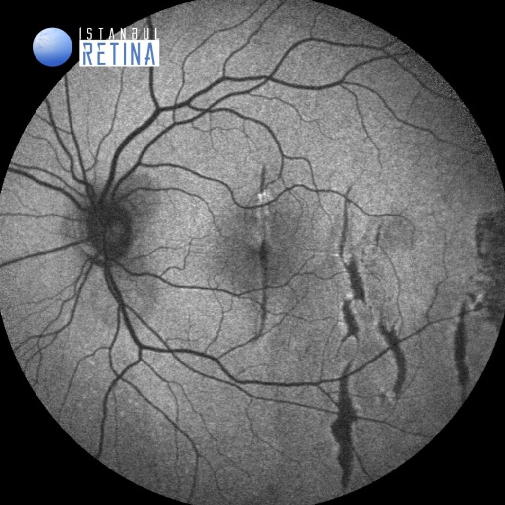
Fundus autofluorescence imaging demonstrated hypo-autofluorescence areas representing the choroidal rupture (Figure 2).
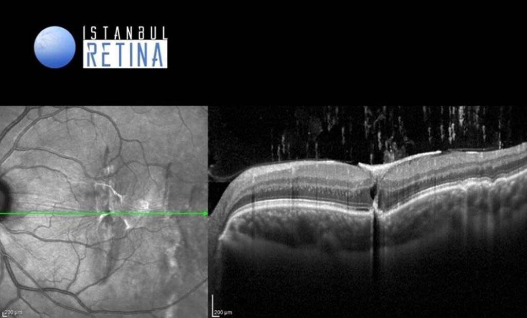
Spectral–domain OCT imaging revealed deffective retinal pigment epithelium-Bruch’s membrane complex and epiretinal membrane (Figure 3).
Diagnosis
Choroidal Rupture
A choroidal rupture is a break in the choroid, Bruch membrane, and the retinal pigment epithelium (RPE). A choroidal rupture usually is a result of a traumatic injury. During a closed globe injury, the eyeball is first mechanically compressed and then rapidly hyperextended. The sclera’s tensile strength resists this compression. The retina is elastic and stretches during such an injury. However, Bruch membrane breaks because it does not have sufficient tensile strength or elasticity. The choriocapillaris is injured and bleeds into the subRPE and/or subretinal space. Such hemorrhage may hide the choroidal rupture initially. Over days, the blood clears and a whitish/yellowish, curvilinear, crescent-shaped subretinal streak is visible. It is usually concentric to the optic nerve. In rare cases, the choroidal rupture may be oriented radially. There may be one or more choroidal ruptures present. Over time, choroidal neovascularization (CNV) can develop. If the rupture or CNV does not involve the foveal center, vision may not be affected.
Differential Diagnosis
Lacquer cracks in an eye with high myopia, angioid streaks
Treatment
Observation is recommended with Amsler grid monitoring. However, in the presence of CNV, anti-vascular endothelial growth factor injections can be used.
References:
Lupidi M, Muzi A, Castellucci G, et al. The choroidal rupture: current concepts and insights. Surv Ophthalmol. 2021;66:761-770. https://pubmed.ncbi.nlm.nih.gov/33545177/