Medical History:
A 50-year-old female patient presented with complaint of vision loss in the left eye.
Diabetes mellitus (-)
Systemic hypertension (-)
Family history (-)
Smoking (-)
Trauma (-)
Examination Findings
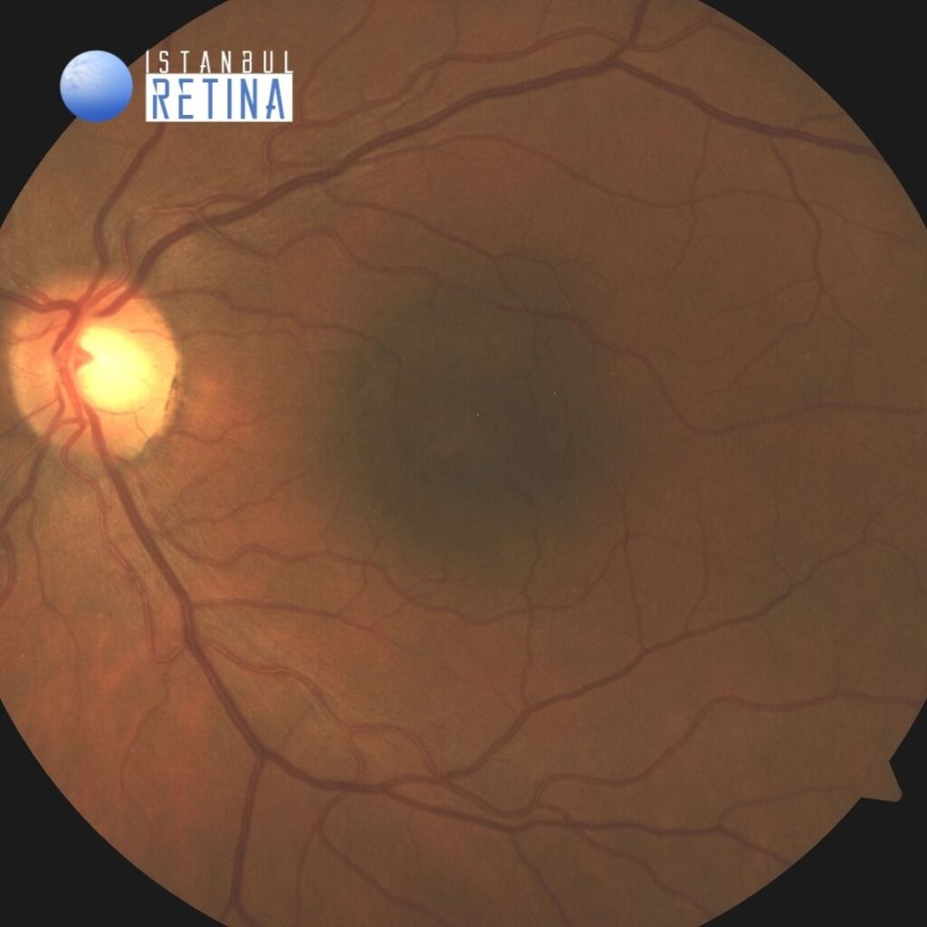
Best corrected visual acuity was 10/10 in the right eye and 5/10 in the left eye. Intraocular pressure was 14 mmHg in the right eye and 12 in the lefte ye. Anterior segment examination was unremarkable. Fundus evaluation of the right eye was normal. In the patient’s left eye, a subfoveal pigmented choroidal nevus was seen, measuring 2 disc diameters in basal diameter (Figure 1).
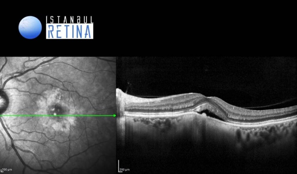
On OCT, there is gradual transition between the hyperreflective inner choroid and hyporeflective outer choroid. Overlying subretinal fluid was detected. The overlying retinal pigment epithelium showed slight irregularity, and the foveal photoreceptors appeared thickened and shaggy (Figure 2).
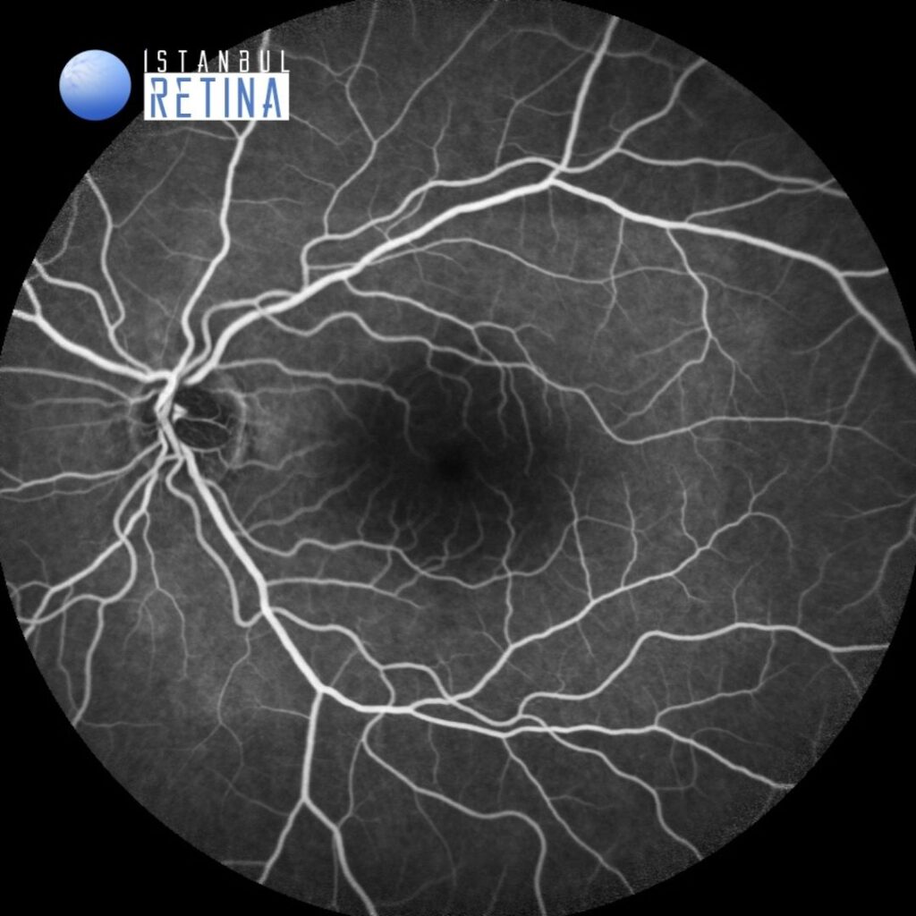
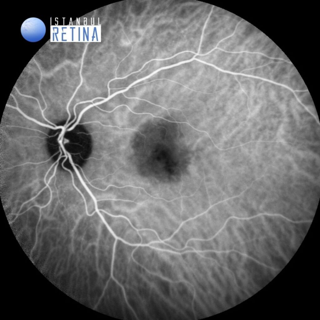
Fluorescein angiography (Figure 3) and indocyanine green angiography (Figure 4) demonstrated hypofluorescence overlying the nevus.
Diagnosis
Subretinal Fluid Associated with Choroidal Nevus
Choroidal nevus is a benign melanocytic lesion of the eye. Choroidal nevus can produce symptoms of visual loss or floaters related to subfoveal location, overlying retinal edema or photoreceptor loss, choroidal neovascularization (CNV), and related subretinal fluid. On OCT, the choroidal findings in nevi are limited to the anterior surface and include hyporeflectivity in 62%, isoreflectivity in 29%, and hyperreflectivity in 9%. Anterior choroidal reflectivity is affected by overlying retinal pigment epithelium alterations and the amount of pigmentation.
Treatment
Choroidal nevus is a benign, typically asymptomatic condition that requires only observation. There are cases in which subretinal fluid overlying a bland subfoveal or perifoveal nevus leads to visual loss and requires medical intervention. Treatment options in such cases, particularly those in which the nevus shows a lack of other risk factors for melanoma, include laser photocoagulation, transpupillary thermotherapy, PDT, and anti-VEGF therapy.
References:
1. Say EA, Shah SU, Ferenczy S, Shields CL. Optical coherence tomography of retinal and choroidal tumors. J Ophthalmol. 2012;2012:385058. https://pubmed.ncbi.nlm.nih.gov/23008756/
2. Pointdujour-Lim R, Mashayekhi A, Shields JA, Shields CL. Photodynamic therapy for choroidal nevus wıth subfoveal fluıd. Retina. 2017;37:718-723. https://pubmed.ncbi.nlm.nih.gov/27433882/