Medical History:
A 60-year-old female patient presented with complaint of floaters in the right eye.
Diabetes mellitus (-)
Systemic hypertension (-)
Family history (-)
Smoking (-)
Trauma (-)
Examination Findings
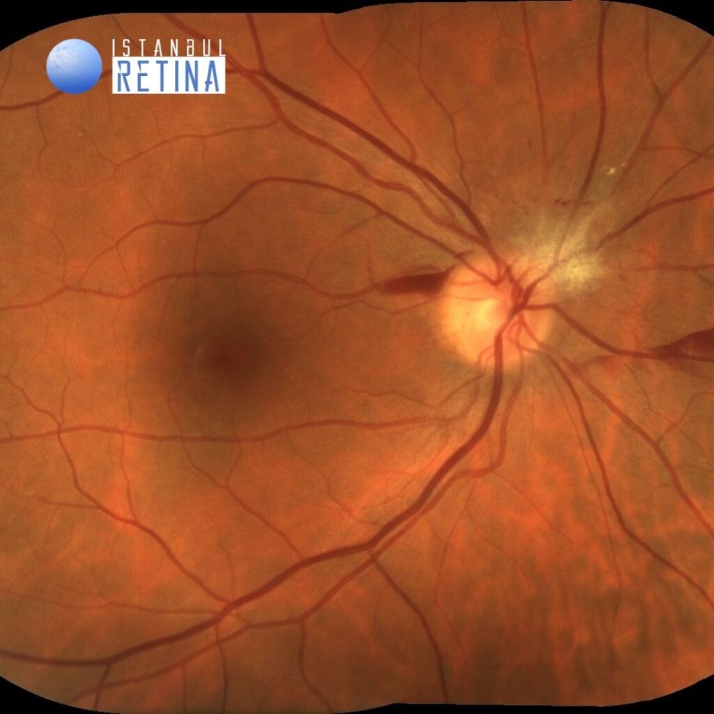
Best corrected visual acuity was 10/10 in the right eye and 10/10 in the left eye. Intraocular pressure was 17 mmHg in the right eye and 17 in the lefte ye. Anterior segment examination was unremarkable. Fundus examination revealed preretinal hemorrhages and a hypopigmented lesion located near the superonasal optic disc margin in the right eye. The left fundus was normal (Figure 1).
Optic coherence tomography (OCT) scan through lesion demonstrated full intraretinal extent of the lesion (Figure 2).
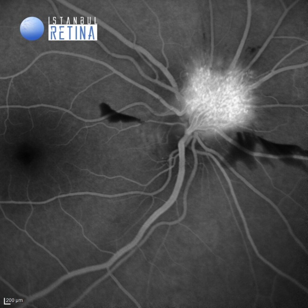
Early phase fluorescein angiography showed a well-circumscribed hyperfluorescent vascular structure (Figure 3).

Fluorescein angiography revealed leakage in the late phases (Figure 4).
Diagnosis
Exophytic Juxtapapillary Retinal Capillary Hemangioma
Juxtapapillary retinal capillary hemangiomas are vascular hamartomas that occur on or adjacent to the optic nerve head. Juxtapapillary retinal capillary hemangiomas may occur sporadically or in association with von Hippel–Lindau disease. Three distinct growth types of juxtapapillary retinal capillary hemangiomas have been described, including the endophytic, exophytic, and sessile forms. Mostretinal capillary hemangiomas present as endophytic, red-orange, nodular retinalmasses growing from the inner retinal surface in the direction of the vitreous cavity. Less commonly, RCH can grow in an exophytic pattern, in the direction of the subretinal space, in whichcase they often have a more subtle appearance that can be challenging to diagnose.
Differential Diagnosis
Papilledema, papillitis, choroidal neovascular membrane.
Treatment
Because of the location of these hemangiomas, treatment is very complex. These lesions should be treated if vision is reduced or if there is lesion progression. Several treatments have been proposed, such as laser photocoagulation, brachytherapy, transpupillary thermotherapy, photodynamic therapy, and surgical excision. None of these treatments has proven to be particularly effective in inducing regression.
References:
1. Saitta A, Nicolai M, Giovannini A, Mariotti C. Juxtapapillary retinal capillary hemangioma: new therapeutic strategies. Med Hypothesis Discov Innov Ophthalmol 2014;3:71-5. https://pubmed.ncbi.nlm.nih.gov/25741522/
2. Custo Greig EP, Duker JS. Retinal hemangioblastoma vascular detail elucidated on swept source optical coherence tomography angiography. Am J Ophthalmol Case Rep 2020;21:101005. https://pubmed.ncbi.nlm.nih.gov/33385098/
3. Gass JD, Braunstein R. Sessile and exophytic capillary angiomas of the juxtapapillary retina and optic nerve head. Arch Ophthalmol 1980;98:1790-7. https://pubmed.ncbi.nlm.nih.gov/7425905/
4. Russell JF, Villegas VM, Schwartz SG, Weng CY, Davis JL, Flynn HW Jr, Harbour JW. Multimodal Imaging in the Diagnosis of Exophytic Juxtapapillary Retinal Capillary Hemangioblastoma. Am J Ophthalmol 2021;225:128-136. https://pubmed.ncbi.nlm.nih.gov/33450232/