Medical History:
A 60-year-old male patient presented to our clinic with a three-day history of vision loss his right eye.
Diabetes mellitus (-)
Systemic hypertension (-)
Family history (-)
Smoking (-)
Trauma (-)
Examination Findings
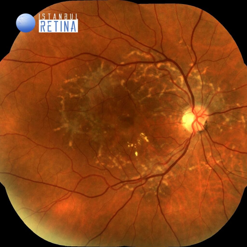
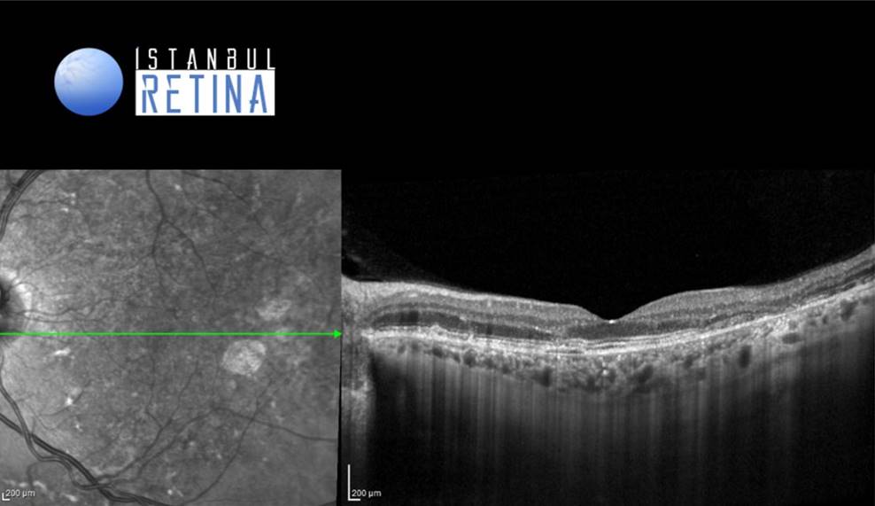
Best corrected visual acuity was 6/10 in the right eye and 9/10 in the left eye. Intraocular pressure was 15 mmHg in both eyes. Anterior segment examination was unremarkable. Funduscopic examination revealed multiple yellowish flecks over the posterior pole and hard exudates inferonasal to the fovea in the right eye (Figure 1).

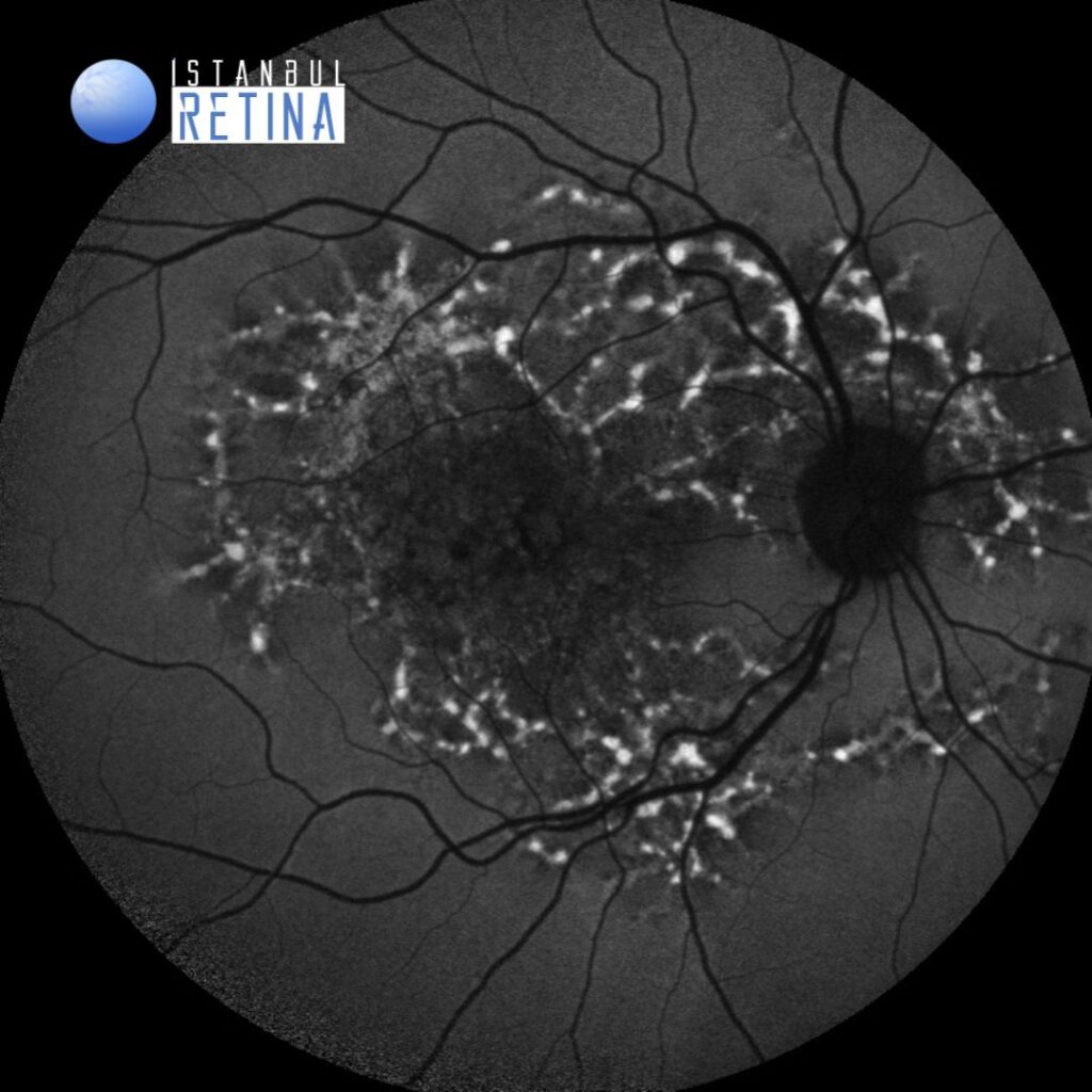
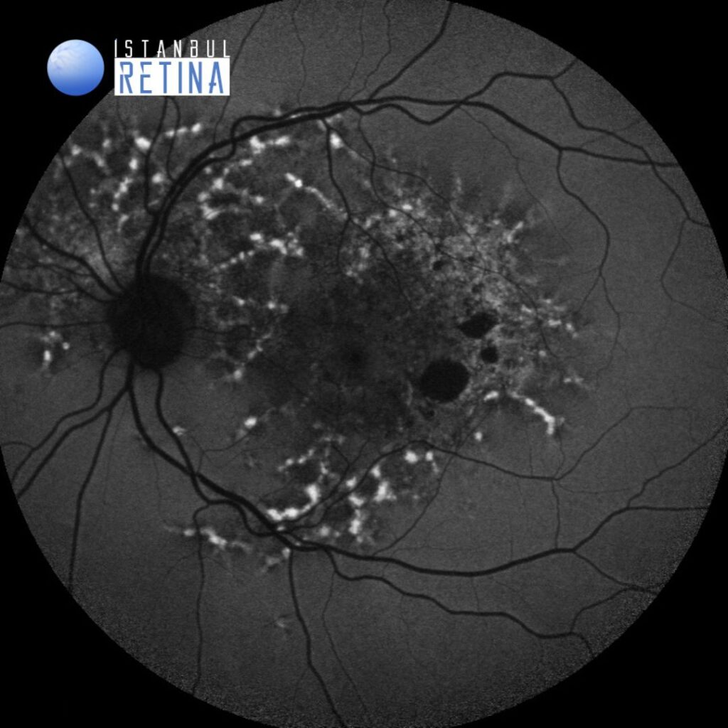
Autofluorescence image of both eyes showed multiple hyperautofluorescent lesions corresponding to flecks (Figure 2).
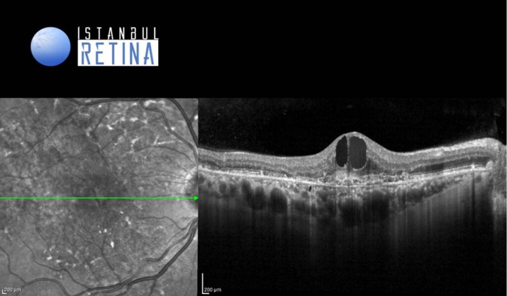
Spectral domain optical coherence tomography of the right eye showed subretinal hyperreflective material and intraretinal cystic spaces (Figure 3).
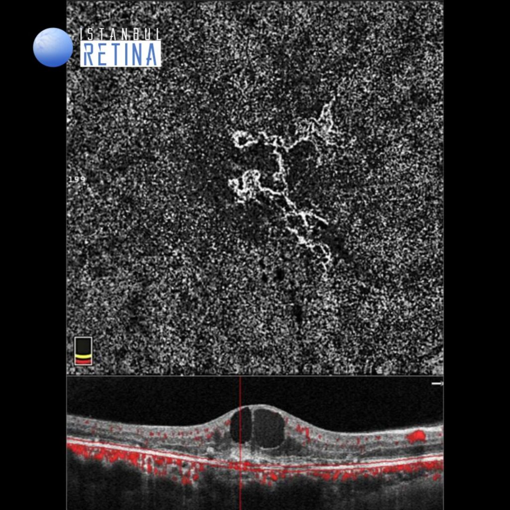
Optical coherence tomography angiography of the right eye revealed branched vascular network (Figure 4).
Diagnosis
Choroidal neovascular membrane secondary to pattern dystrophy simulating fundus flavimaculatus
Pattern dystrophies are a group of inherited disorders of the retinal pigment epithelium. Pattern dystrophies are divided into 5 types based on the pattern of pigment distribution: adult-onset foveo macular vitelliform dystrophy, butterfly-shaped pigment dystrophy, reticular dystrophy of the retinal pigment epithelium, pattern dystrophy simulating fundus flavimaculatus, and fundus pulverulentus. Pattern dystrophies usually have a good visual prognosis till later life, but can be rarely associated with other complications like choroidal neovascular membrane. Choroidal neovascular membrane in pattern dystrophy simulating fundus flavimaculatus is extremely rare.
Differential Diagnosis
Neovascular age related macular degeneration, pachychoroid neovasculopathy, neovascularization secondary to angioid streaks, neovascularization secondary to Best’s vitelliform macular dystrophy
Treatment
Anti-VEGF injections have been shown to improve both anatomical and functional outcomes in patients with choroidal neovascular membrane secondary to pattern dystrophies.
References:
Nangia P, Shah D, Saurabh K, Roy R. Efficacy of anti-VEGF in the treatment of choroidal neovascular membrane secondary to pattern dystrophy simulating fundus flavimaculatus. GMS Ophthalmol Cases. 2019;9:21. https://www.ncbi.nlm.nih.gov/pmc/articles/PMC6637455/