Medical History:
A 27-year-old female patient presented with complaint of blurry vision in the right eye for the past 3 weeks.
Diabetes mellitus (-)
Systemic hypertension (-)
Family history (-)
Smoking (-)
Trauma (-)
Examination Findings
Small yellowish papules coalescing into larger plaques in the back of the patient’s neck were observed.
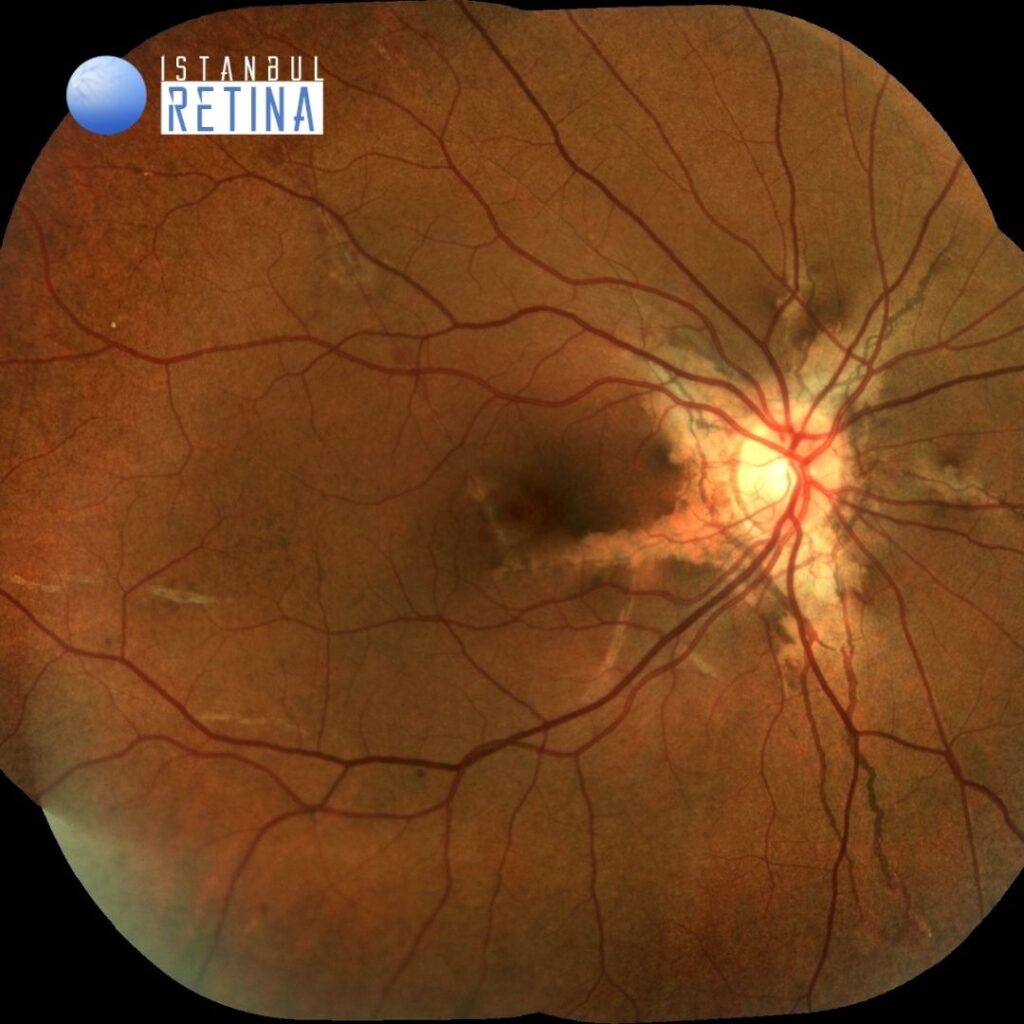
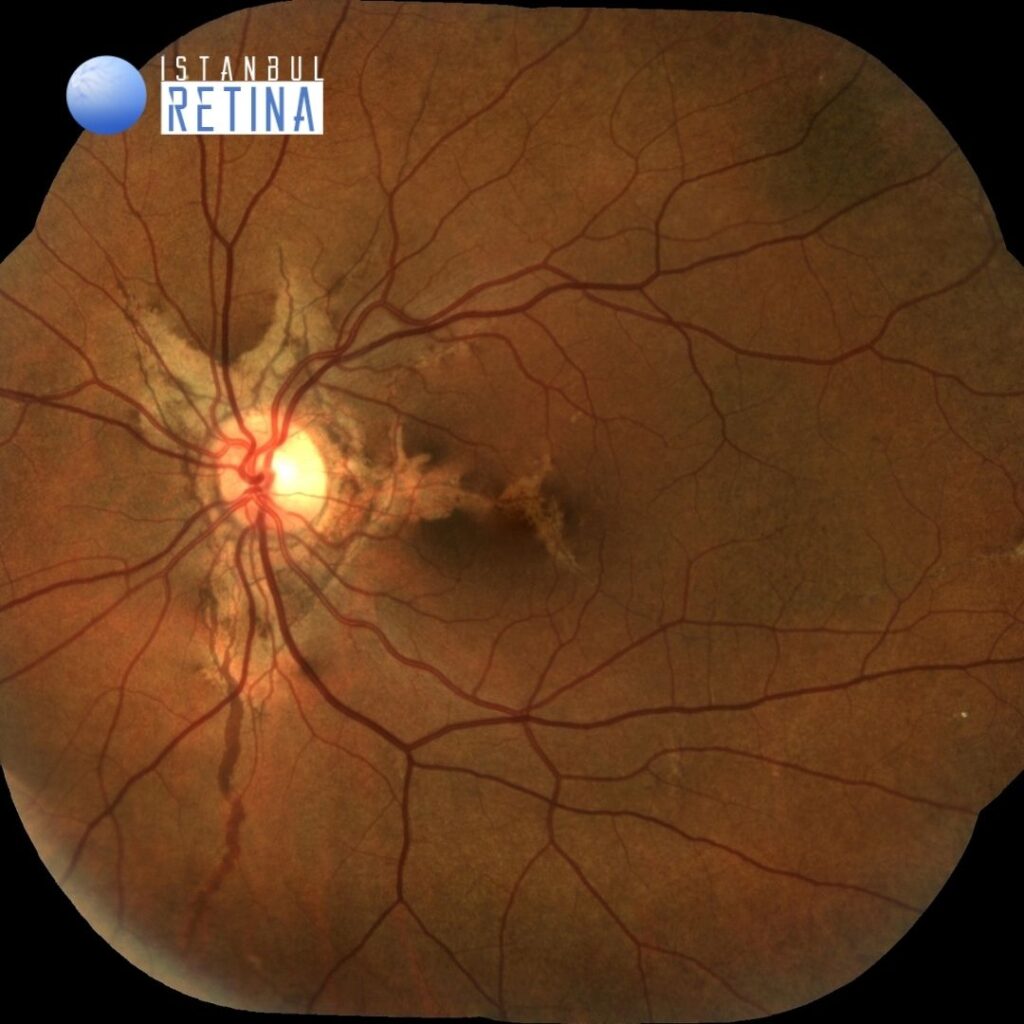
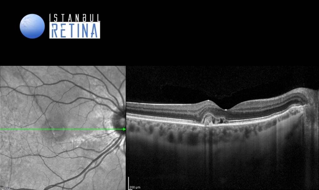
Best corrected visual acuity was 6/10 in the right eye and 10/10 in the left eye. Intraocular pressure was 15 mmHg in both eyes. The anterior segment exam was unremarkable in both eyes. Dilated funduscopic examination revealed peau d’orange appearance in the temporal parts of the retina and angioid streaks extending radially from the optic disc and some involving the fovea in both eyes (Figure 1).
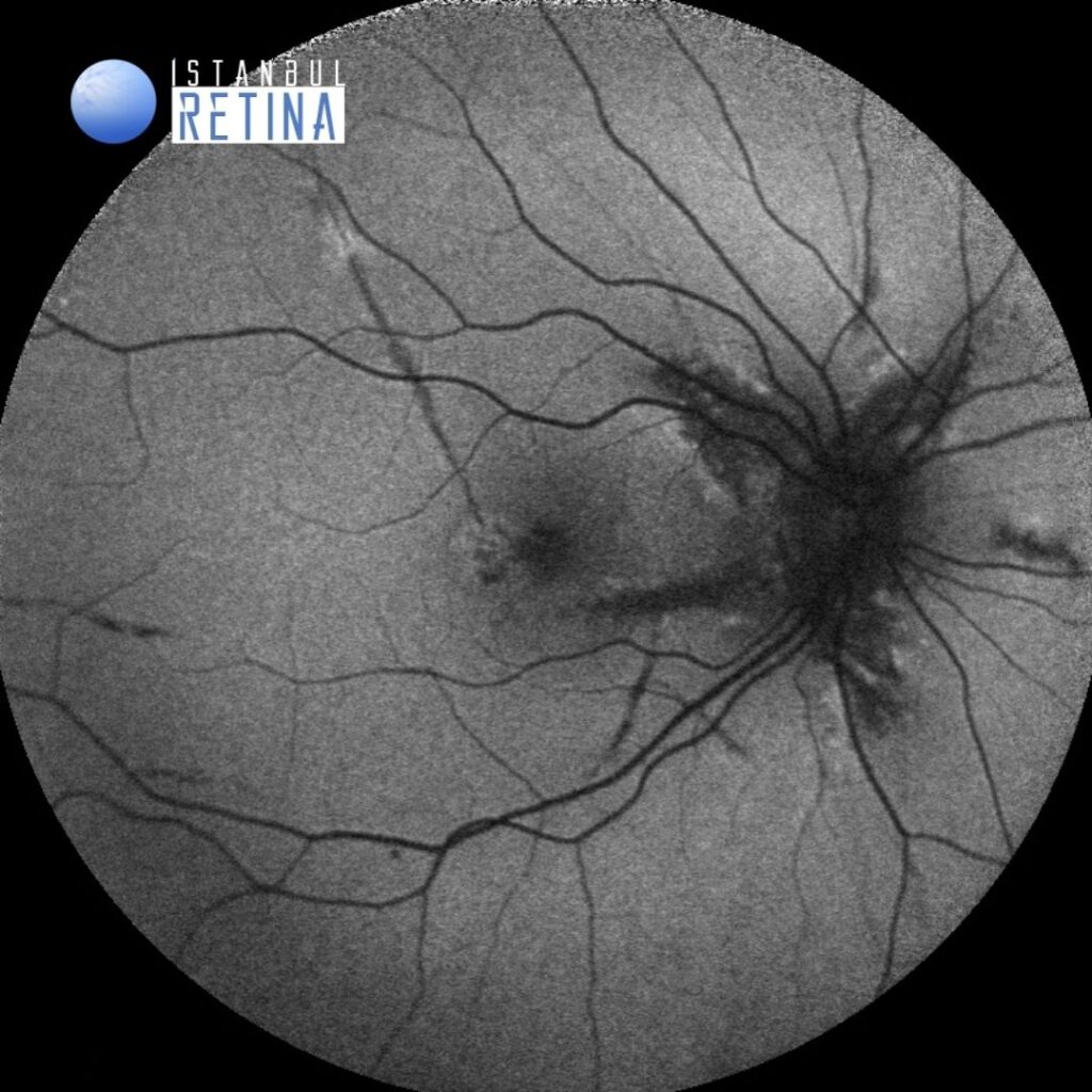
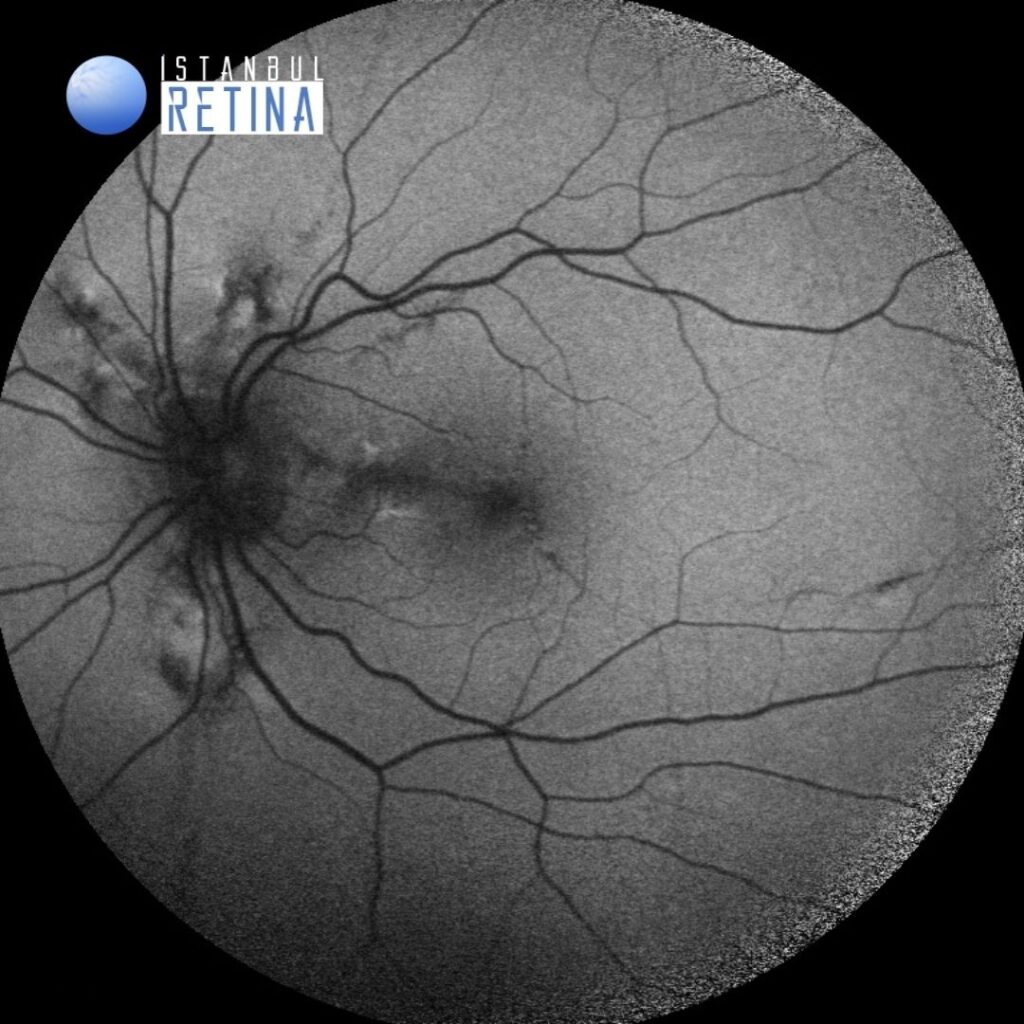
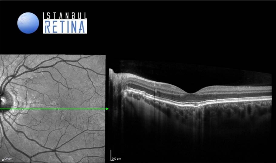
Angioid streaks on fundus autofluorescence appeared as radial hypoautofluorescent fissuresfissures (Figure 2).
SD-OCT revealed revealed subretinal hyperreflective material and subretinal fluid in the right eye (Figure 3).
Diagnosis
Choroidal Neovascularization Associated with Angioid Streaks in Pseudoxanthoma Elasticum
Pseudoxanthoma elasticum is a rare genetic, multisystemic disease which affects primarily the skin, the eyes, and the vascular system. The prevalence of pseudoxanthoma elasticum is approximately 1:50000-200000. Pseudoxanthoma elasticum is characterized by progressive calcification and degeneration in elastic fibrils and is associated with the mutation in the gene encoding the ABCC6 (MRP6) transmembrane transport protein. Ocular abnormalities or complications are angioid streaks, peau d’orange appearance, comet lesions, and choroidal neovascularization.
Generally, the first signs of pseudoxanthoma elasticum are the asymptomatic cutaneous features and angioid streaks are the most frequent ocular findings. Angioid streaks are gray-white lines with rupture of the thickened elastic fibers of Bruch’s membrane extending radially from the optic disc and occur in 80% of patients with pseudoxanthoma elasticum. Choroidal neovascularization occurs in 42–84% of patients with pseudoxanthoma elasticum.
Differential Diagnosis
Angioid streaks are not the pathognomonic sign of pseudoxanthoma elasticum and may occur due to Paget’s disease, Marfan syndrome, Ehler-Danlos syndrom, sickle-cell anemia, and beta thalassemia.
Treatment
To date, there is no effective treatment for pseudoxanthoma elasticum or specific therapies for the systemic complications of this disease. Nevertheless, intravitreal anti-VEGF injections have been performed to increase visual acuity in pseudoxanthoma elasticum complicated with choroidal neovascularization.
References:
- Mirza E, Karanfil FC, Mirza GD, Choroidal neovascularization associated with angioid streaks in a patient with pseudoxanthoma elasticum. Biomed J Sci & Tech Res. 2018 https://biomedres.us/fulltexts/BJSTR.MS.ID.001995.php
- Hocaoglu M, Karacorlu M, Sayman Muslubas I, Arf S, Ozdemir H, Uysal O. Visual impairment in pseudoxanthoma elasticum: a survey of 53 patients from Turkey. Eur J Ophthalmol. 2016;26(5):449-53. https://pubmed.ncbi.nlm.nih.gov/27150937/