Medical History:
A 75-year-old female patient presented with complaint of gradually progressive blurring of vision in the left eye for the past 3 months.
Diabetes mellitus (+)
Systemic hypertension (+)
Family history (-)
Smoking (-)
Trauma (-)
Examination Findings
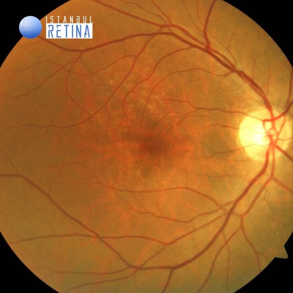
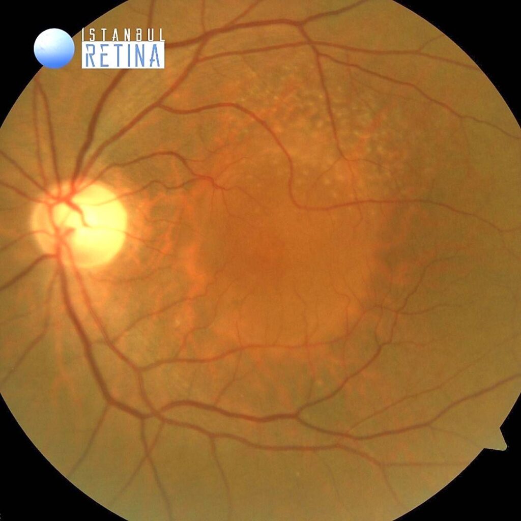
Best corrected visual acuity was 9/10 in the right eye and 3/10 in the left eye. Intraocular pressure was 17 mmHg in both eyes. Anterior segment examination showed nuclear sclerosis in both eyes. Funduscopic examination revealed bilateral reticular pseudodrusen and dome-shaped elevation of the retinal pigment epithelium (RPE) with sharp borders in the left eye (Figure 1).
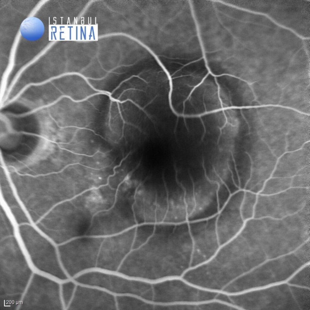
Fluorescein angiography (FA) showed early hyperfluorescence and gradual pooling. Presence of a hyperfluorescent notch at the nasal edge of the retinal pigment epithelial detachment (PED) was observed (Figure 2).
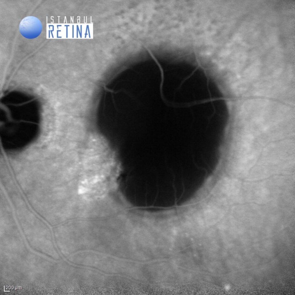
Indocyanine angiography (ICGA) showed a serous PED as an area of hypofluorescence and hyperfluorescent plaque associated with the choroidal neovascularization (CNV) (Figure 3).
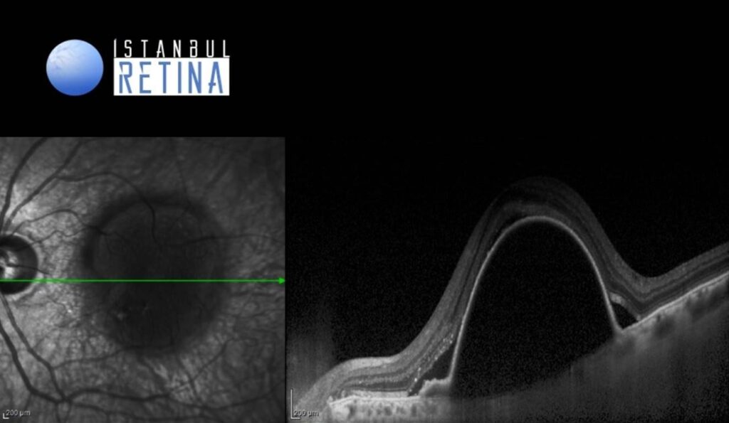
SD-OCT revealed dome shaped elevation of the RPE over a hypo-reflective space and subretinal fluid (Figure 4).
Diagnosis
Vascularized serous pigment epithelial detachment in age-related macular degeneration
PEDs in AMD are usually classified as serous PED, fibrovascular PED, and drusenoid PED. Serous PED may occur with (vascularized PED) or, without CNV (nonvascularized PED). Most of the serous PEDs are vascularized. ICGA imaging revealed that CNV is observed in 96% of patients with serous PED.
On fundoscopy, serous PEDs may have a translucent or an orange-yellowish appearance. On FA, they exhibit early hyperfluorescence and gradual pooling. The presence of a hyperfluorescent notch at the edge of the PED is a strong sign of the presence of CNV. A serous PED is typically seen as an area of hypofluorescence on ICGA. In contrast, a vascularized serous PED will have a hyperfluorescent notch or plaque associated with the CNV.
Differential Diagnosis
Nonvascularized serous PED, fibrovascular PED, drusenoid PED
Treatment
Currently, serous PEDs associated with neovascular AMD are treated with intravitreal anti-VEGF agents alone or in combination with photodynamic therapy.
References:
- Yannuzzi LA, Hope-Ross M, Slakter JS, et al. Analysis of vascularized pigment epithelial detachments using indocyanine green videoangiography. Retina. 1994;14:99-113. https://pubmed.ncbi.nlm.nih.gov/7518607/
- Querques G, Pastore MR, Khlifi H, et al. Integrated Imaging of Avascular Serous Pigment Epithelium Detachment in Age-related Macular Degeneration. Ophthalmology @ Point of Care. January 2017. https://journals.sagepub.com/doi/full/10.5301/oapoc.0000009
- Karampelas M, Malamos P, Petrou P, et al. Retinal Pigment Epithelial Detachment in Age-Related Macular Degeneration. Ophthalmol Ther. 2020;9:739-756. https://www.ncbi.nlm.nih.gov/pmc/articles/PMC7708599/