Medical History:
A 34-year-old male patient presented with complaint of blurry vision in her left eye for 3 months.
Diabetes mellitus (-)
Systemic hypertension (-)
Family history (-)
Smoking (-)
Trauma (-)
Examination Findings
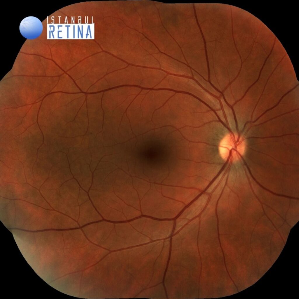
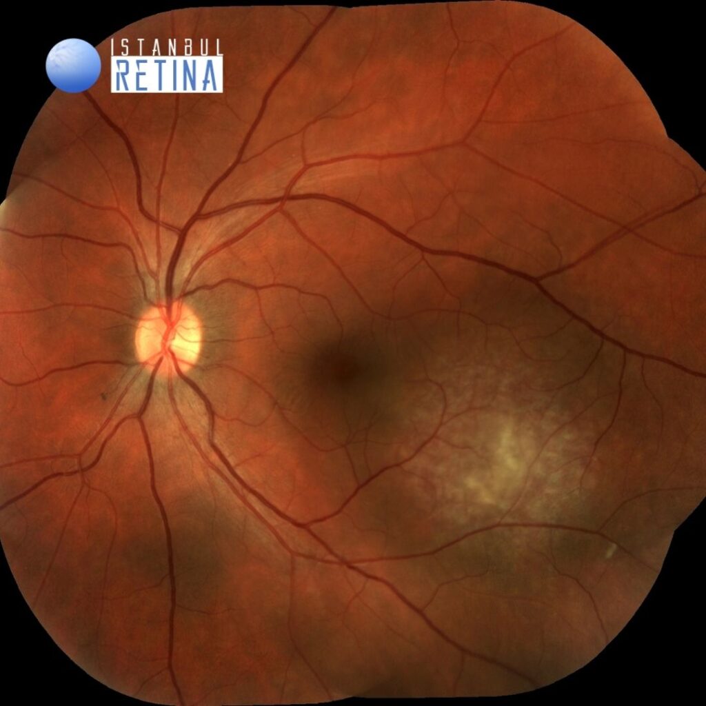
Best corrected visual acuity was 10/10 in the right eye and 4/10 in the left eye. Intraocular pressure was 15 mmHg in both eyes. Anterior segment examination was unremarkable in both eyes. Funduscopic examination was unremarkable in the right eye (Figure 1A). The funduscopic examination in the left eye showed orange, round choroidal tumor located inferior-temporal to the fovea (Figure 1B).
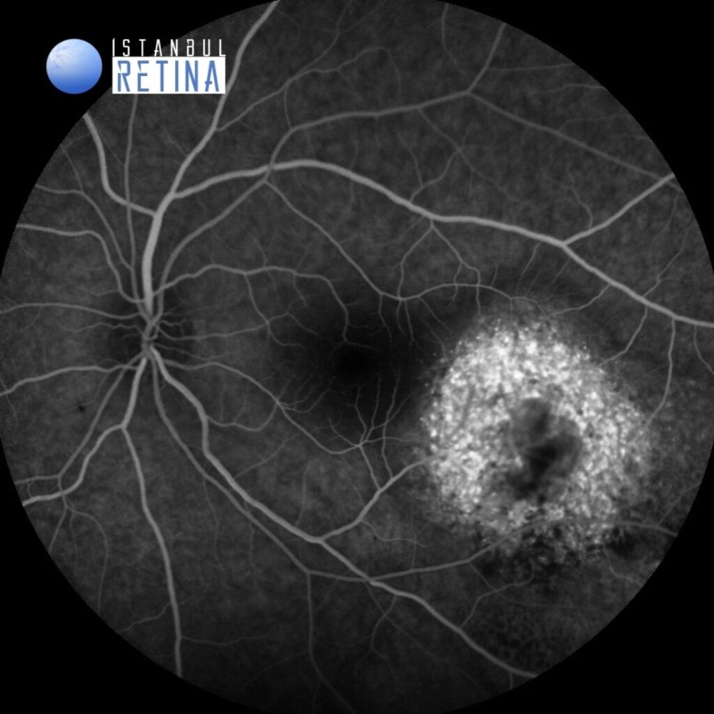
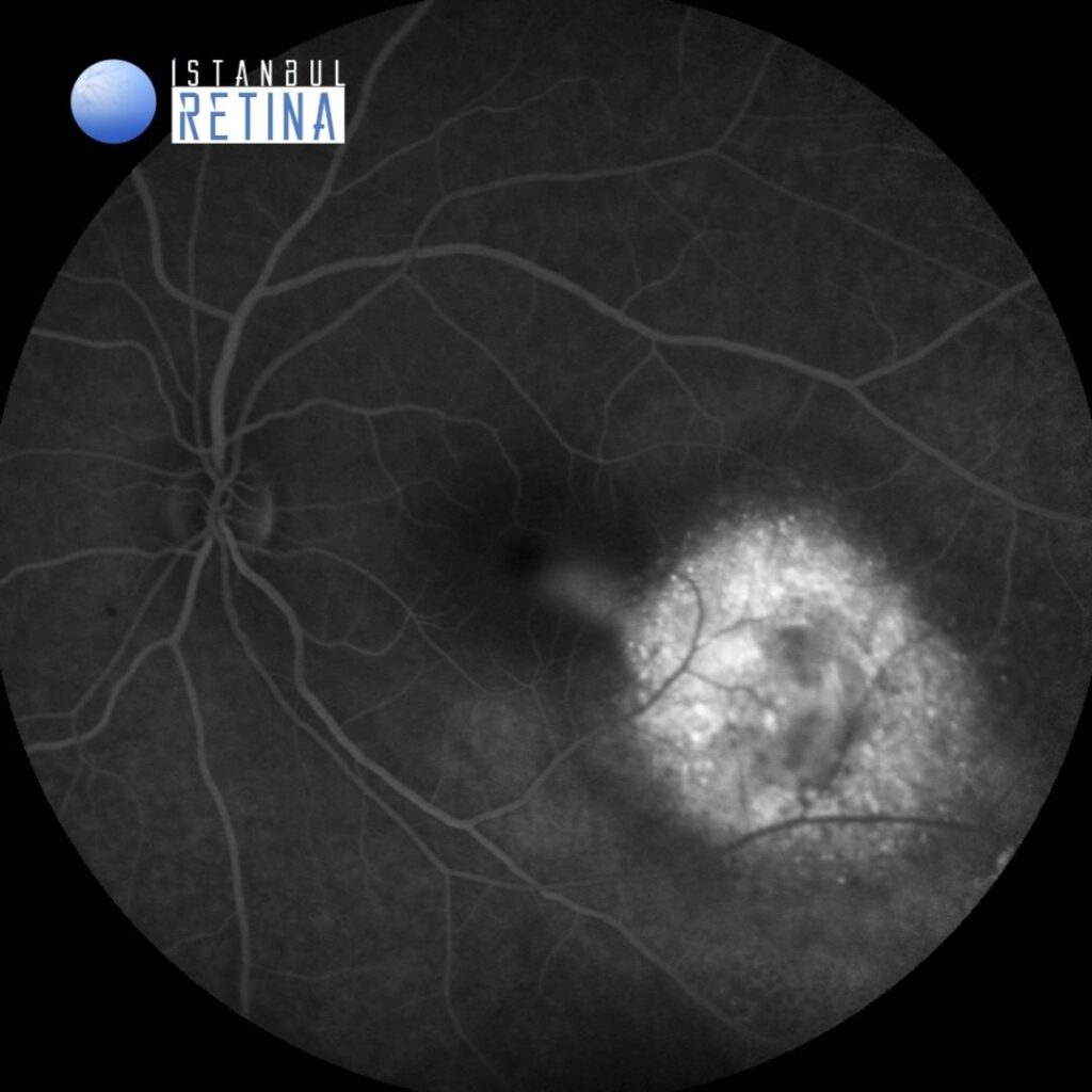
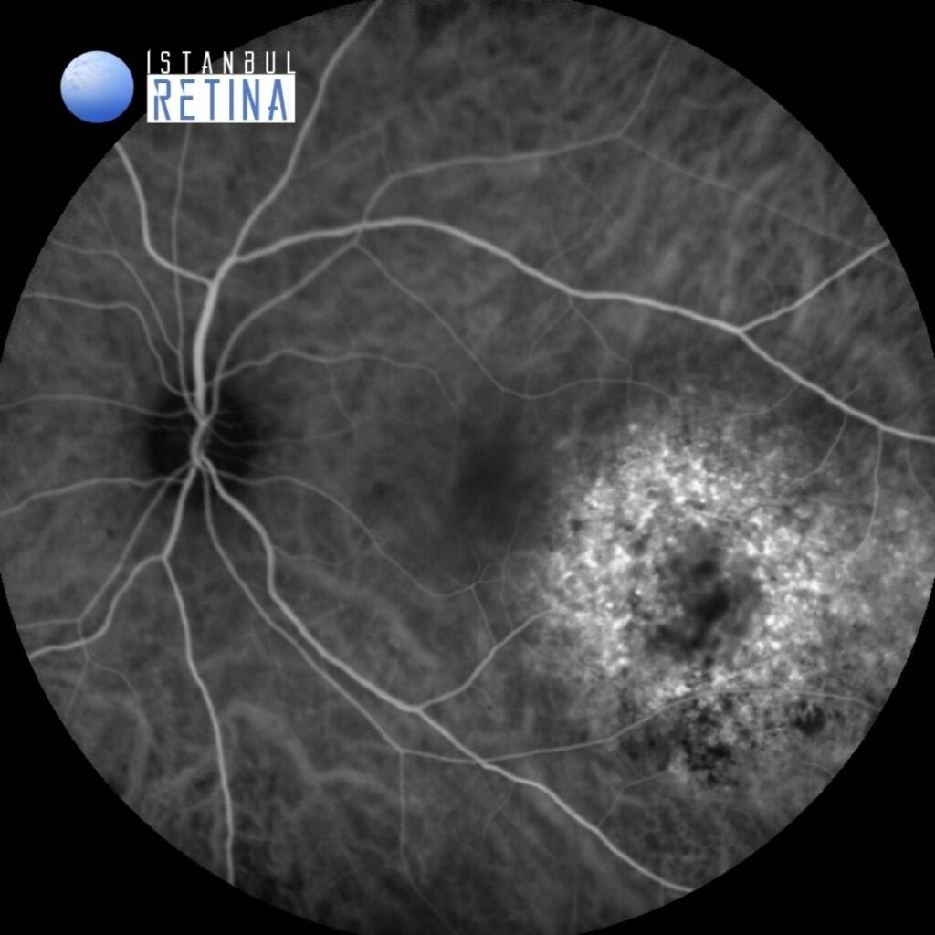
Fluorescein angiography demonstrated a ring of early hyperflourescence (Figure 1A) that increased through late phase (Figure 2B).
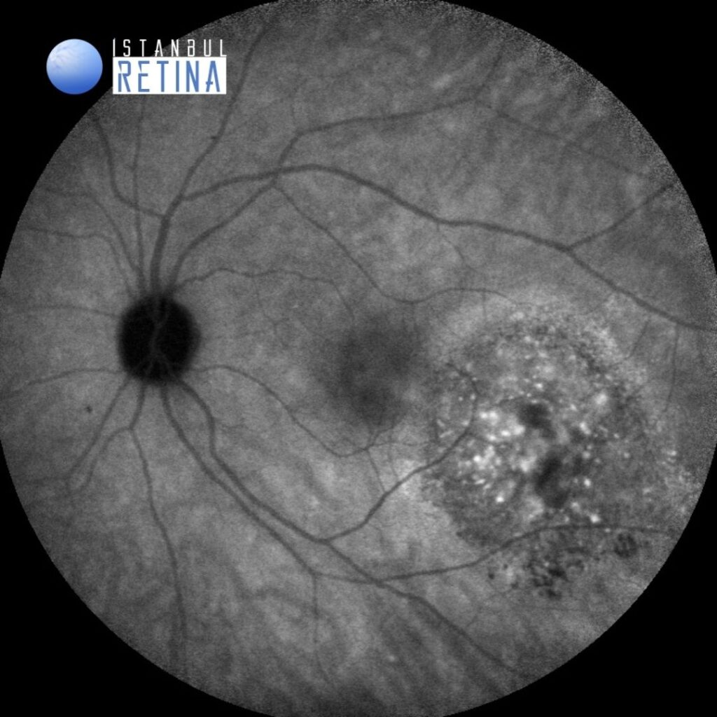
Indocyanine green angiography showed extreme hyperflourescence in the early stages (Figure 3A) which decreased in the late stages (Figure 3B).
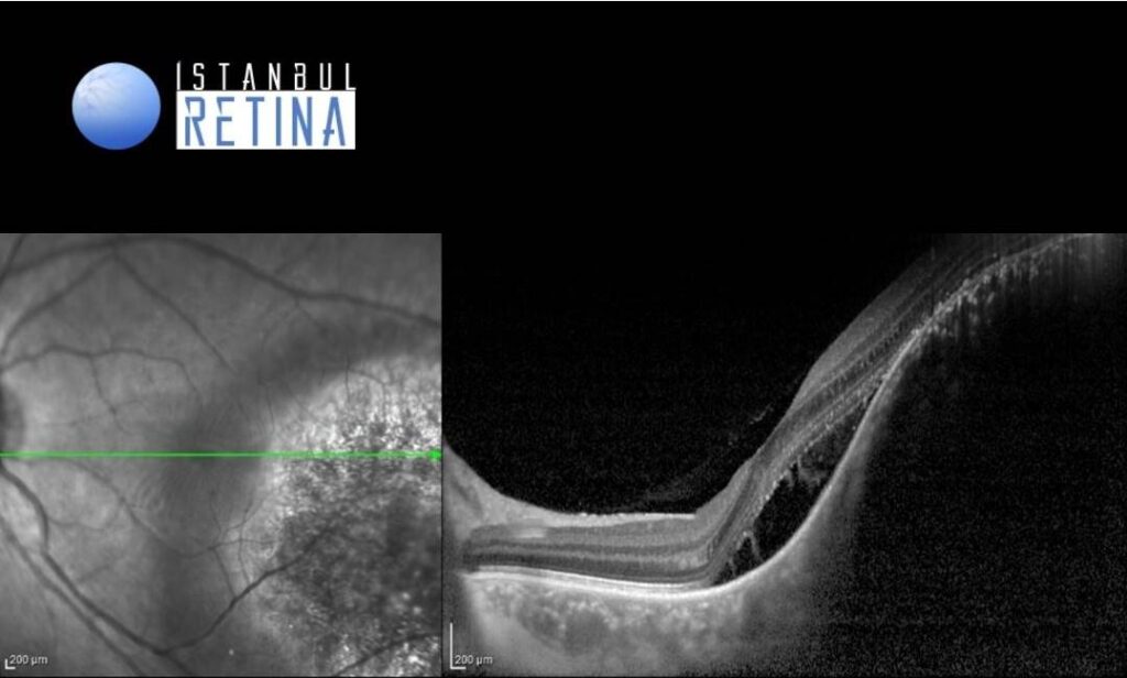
SD-OCT revealed dome-shaped lesion involving the choroid and subretinal fluid (Figure 4).
Diagnosis
Circumscribed Choroidal Hemangioma
Choroidal hemangioma is a benign hamartomatous disorder that is almost always isolated and nonsyndromic, and diagnosed between the second and fourth decade of life. Most circumscribed choroidal hemangiomas are noted first when they produce visual symptoms caused by accumulation of serous subretinal fluid, degenerative changes in the macula or both.
Circumscribed choroidal hemangiomas are noted as reddish-orange, round to oval choroidal tumor located largely in the posterior fundus. Fluorescein angiography typically reveals very early hyperfluorescence of larger-caliber choroidal blood vessels. By late frames, fluorescein commonly stains the entire lesion and any associated subretinal fluid. Indocyanine green angiography typically shows filling of the intralesional vascular channels, intense hyperflourescence of the lesion by the intermediate frames and late “wash-out” of the lesion. Optical coherence tomography can document the dome-shaped lesion involving the choroid, subretinal fluid accumulation along with cystic retinal degeneration that can occur overlying the lesion.
Differential Diagnosis
Choroidal nevus, choroidal melanoma, central serous chorioretinopathy, choroidal metastasis, choroidal osteoma, inflammatory choroidal granuloma, nodular posterior scleritis and Harad´s disease.
Treatment
Asymptomatic cases can be observed. Photodynamic therapy (PDT) using verteporfin has been used with considerable success as treatment of small to medium-size circumscribed choroidal hemangiomas. In some cases PDT has been combined with intravitreal anti-vascular endothelial growth factor drug therapy to treat eyes with a large amount of turbid subretinal fluid. In patients who have an extremely thick choroidal hemangioma, extensive non-rhegmatogenous retinal detachment, or a diffuse or circumscribed choroidal hemangioma that is refractory to PDT, low-dose ocular irradiation appears to be an effective therapeutic option.
References:
1.https://eyewiki.aao.org/Intraocular_Vascular_Tumors#Choroidal_hemangioma. https://eyewiki.aao.org/Intraocular_Vascular_Tumors#Choroidal_hemangioma
2. Berry M, Lucas LJ. Circumscribed choroidal hemangioma: A case report and literature review. J Optom. 2017;10(2):79-83. https://pubmed.ncbi.nlm.nih.gov/26872405/