Medical History:
A 25-year-old female patient presented with complaint of gradually progressive blurring of vision in both eyes for the past 5 years.
Diabetes mellitus (-)
Systemic hypertension (-)
Family history (-)
Smoking (-)
Trauma (-)
Examination Findings
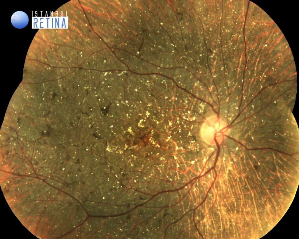
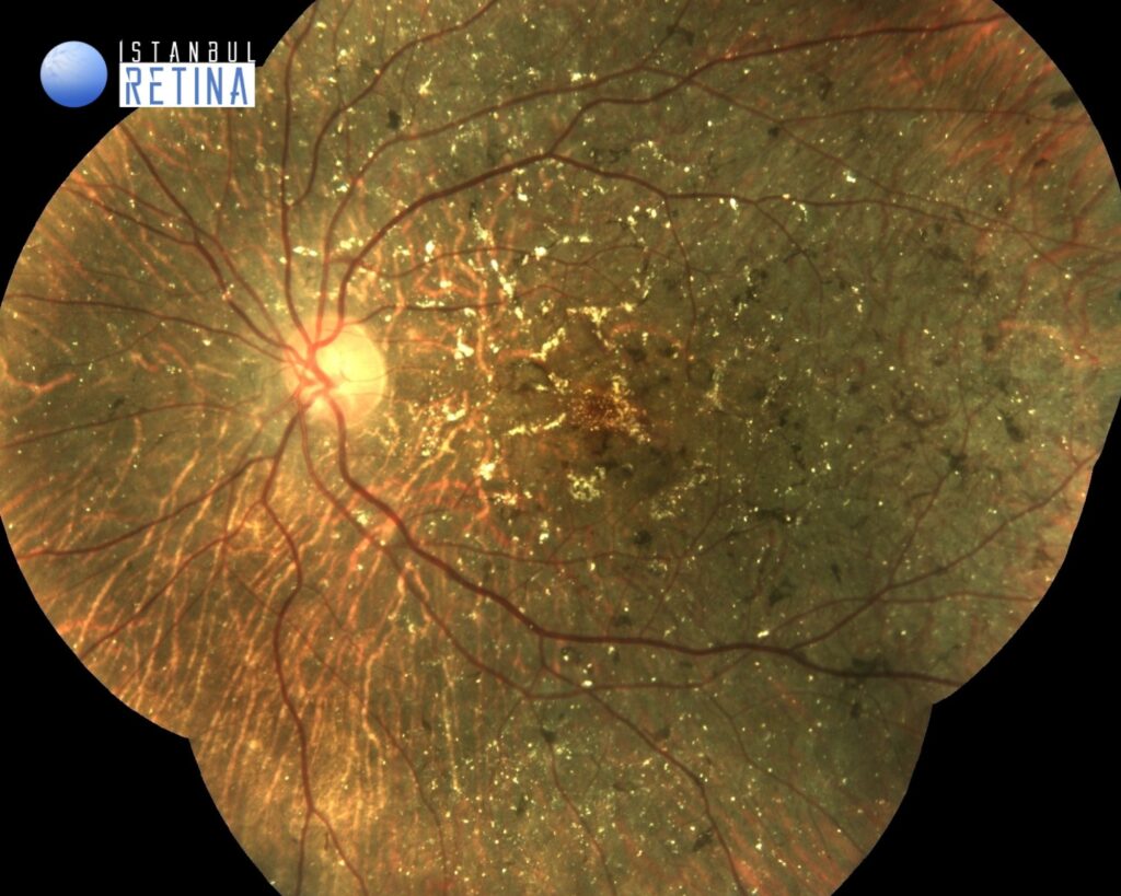
Best corrected visual acuity was 3/10 in the right eye and 3/10 in the left eye. Intraocular pressure was 15 mmHg in both eyes. Anterior segment examination was unremarkable. Funduscopic examination revealed bilateral multiple yellow-white crystalline deposits in the retina, atrophy of the retinal pigment epithelium (RPE), diffuse choriocapillaris atrophy, retinal bone spicule pigmentations and sclerosis of the choroidal vessels (Figure 1).
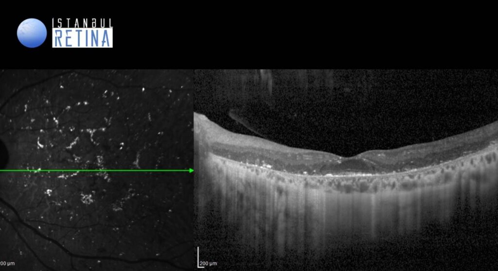
SD-OCT revealed extensive degeneration of the retina and the RPE/choriocapillaris complex, and hyperreflective spots (crystals) located in the RPE/Bruch’s membrane complex (Figure 2).
Diagnosis
Bietti’s Crystalline Dystrophy
Bietti’s Crystalline Dystrophy, is a rare autosomal recessive ocular disease that involves yellow-white crystalline lipid deposits in the retina and sometimes cornea, degeneration of the retinal pigment epithelium and sclerosis of the choroidal vessels. Progression of the disease ultimately results in reduced visual acuity, night blindness, visual field loss, and impaired color vision. Onset of the disease can occur from early teenage years to third decade of life, but can also occur beyond the third decade. As the disease progresses, decreases in peripheral acuity, central acuity or both ultimately results in legal blindness in most patients.
Differential Diagnosis
Retinitis Pigmentosa, Primary hyperoxaluria, Cystinosis, Sjogren-Larsson Syndrome
Treatment
Currently, there are no evidence based surgical treatments or medical treatment specific to Bietti’s crystalline dystrophy available in the existing literature.
References:
- American Academy of Ophthalmology. Bietti crystalline dystrophy. 2019. https://eyewiki.aao.org/Bietti_Crystalline_Dystrophy
- Saatci AO, Doruk HC, Yaman A, Oner FH. Spectral domain optical coherence tomographic findings of bietti crystalline dystrophy. J Ophthalmol. 2014. https://pubmed.ncbi.nlm.nih.gov/25505979/
- Wang W, Chen W, Bai X, Chen L. Multimodal imaging features and genetic findings in Bietti crystalline dystrophy. BMC Ophthalmol. 2020;20:331. https://pubmed.ncbi.nlm.nih.gov/32799831/


