Medical History:
A 60-year-old male patient presented with complaint of blurred vision in both eyes and headache lasting about 1 week. He had a history of coronary artery disease and uncontrolled hypertension.
Diabetes mellitus (-)
Systemic hypertension (+)
Family history (-)
Smoking (+)
Trauma (-)
Examination Findings
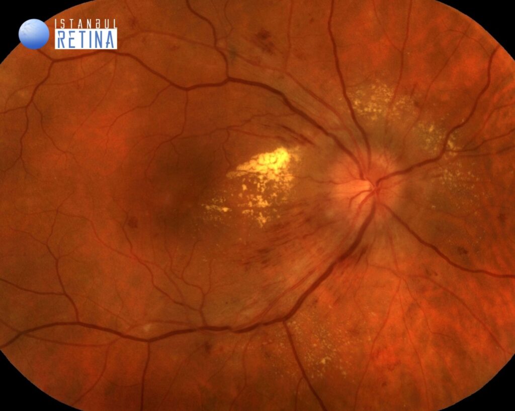
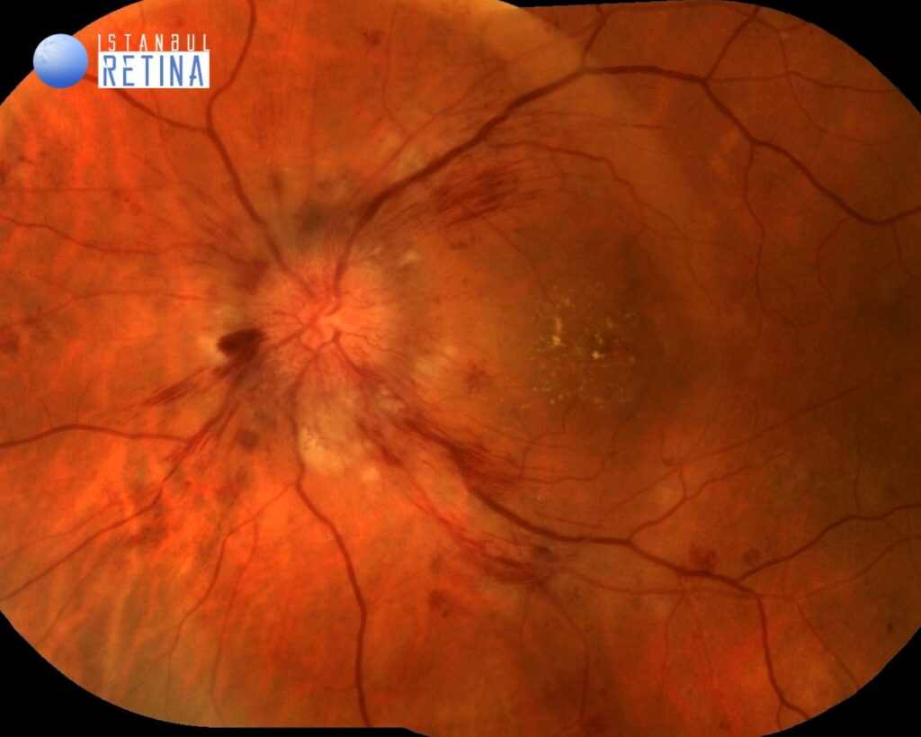
Best corrected visual acuity was 7/10 in the right eye and 3/10 in the left eye. Intraocular pressure was 14 mmHg in both eyes. Anterior segment examination was unremarkable. Funduscopic examination revealed bilateral hard lipid exudates, intraretinal hemorrhages and optic disc swelling, and some cotton wool spots located inferior to the optic nerve in the left eye (Figure 1).
Patients blood pressure reading was 190/140 mmHg.
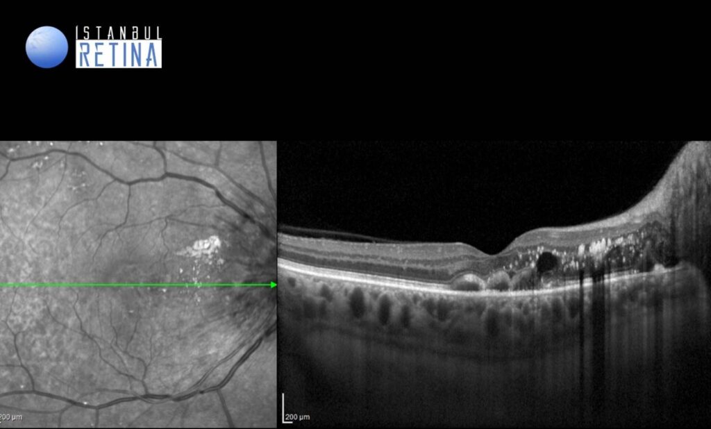
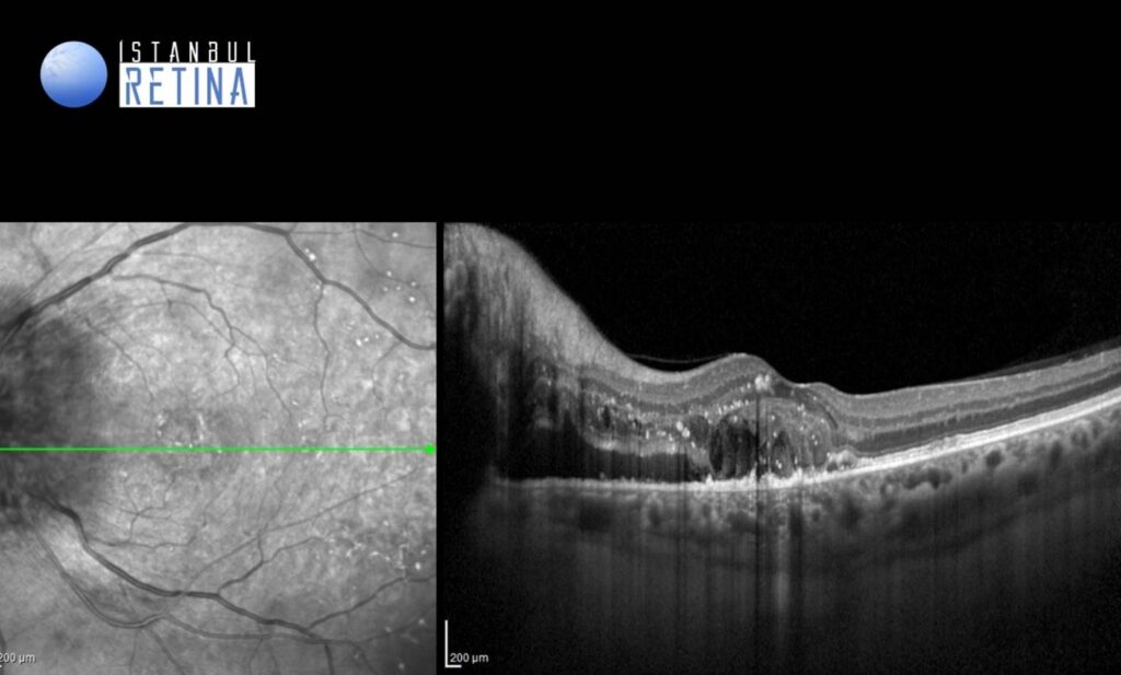
SD-OCT showed intraretinal hyperreflective foci, intraretinal cysts and subretinal fluid located temporal to the optic nerve in both eyes (Figure 2)
Diagnosis
Severe Hypertensive Retinopathy
The vascular changes associated with increased blood pressure are visible in the retina as hypertensive retinopathy. Chronic hypertension leads to structural changes in the vessel wall such as intimal thickening and hyaline degeneration. Thickening of the arterioles leads to compression of the venules where they cross, termed arteriovenous crossing or nicking. Severe hypertension creates focal areas of ischemia of the retinal nerve fiber layer, seen as cotton-wool spots. Breakdown of the blood-retina barrier causes exudation of blood as retinal hemorrhages or exudation of lipids as hard exudates. Very severe hypertension can lead to increased intracranial pressure, causing optic nerve ischemia and optic disc swelling.
Wong and Mitchell Classification of Hypertensive Retinopathy:
Mild: Generalized arteriolar narrowing, focal arteriolar narrowing, arteriovenous nicking, arteriolar wall opacity.
Moderate: Retinal hemorrhage (blot-, dot-, or flame-shaped), microaneurysm, cotton wool spot, hard exudates.
Severe: Moderate retinopathy plus optic disc swelling.
Differential Diagnosis
Diabetic retinopathy, radiation retinopathy, anemia, ocular ischemic syndrome, retinal vein occlusion and neuroretinitis.
Treatment
The treatment for moderate to severe hypertensive retinopathy is primarily focused upon reducing blood pressure.
References:
1. Tsukikawa M, Stacey AW. A Review of Hypertensive Retinopathy and Chorioretinopathy. Clin Optom (Auckl). 2020;12:67-73. https://www.ncbi.nlm.nih.gov/pmc/articles/PMC7211319/
2. Wong TY, Mitchell P. Hypertensive retinopathy. N Engl J Med. 2004;351:2310-7. https://pubmed.ncbi.nlm.nih.gov/15564546/