Medical History:
A 68-year-old male patient with progressive vision loss in both eyes was examined in our clinic.
Diabetes mellitus (-)
Systemic hypertension (+)
Family history (-)
Smoking (+)
Trauma (-)
Examination Findings
Best corrected visual acuity was 7/10 in both eyes. Intraocular pressure was 14 mmHg in both eyes. Anterior segment examination was unremarkable. Funduscopic examination revealed features of a lamellar hole in both eyes (Figure 1).
Fig. 1A
Fig. 1B
In FAF image, increased autofluorescence is found at the fovea (Figure 2).
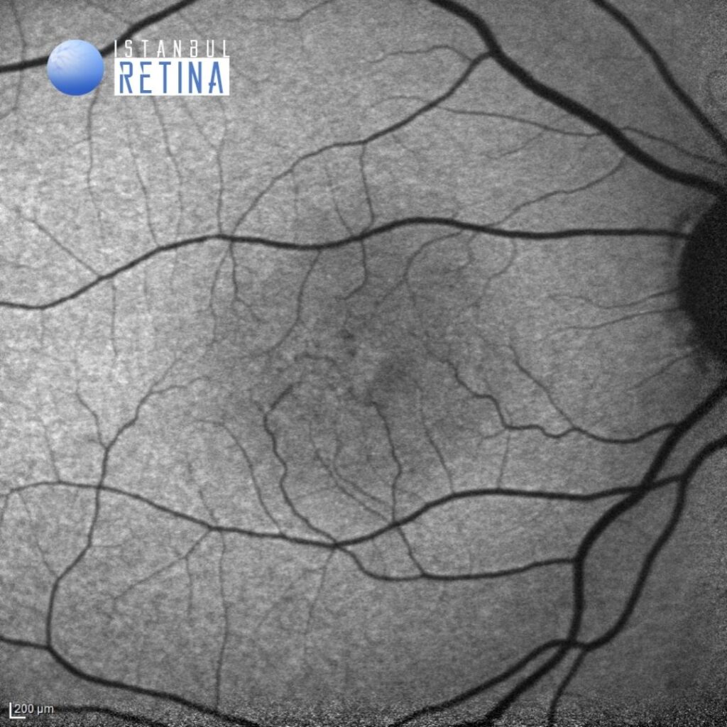
Fig. 2A
Fig. 2B
Abnormalities found in OCT images: Inner and outer retinal cavities at the fovea are accompanied by defect and disruption of external limiting membrane (ELM), ellipsoid zone (EZ) and interdigitation zone (IDZ) at the foveola and temporal parafovea (Figure 3).
Diagnosis
Macular Telangiectasia (MacTel) Type 2
Macular telangiectasia (MacTel) type 2 is an idiopathic bilateral disease that is usually found in middle-aged or older patients. Chronic neurodegenerative processes, vascular inflammation, occlusion and capillary network alterations lead to loss of inner and outer layers of the neurosensory retina and formation of pseudolamellar macular holes.
Imaging findings of MacTel type 2 using spectral domain optical coherence tomography (SD-OCT) include: hyporeflective inner and outer retinal cavities (78%), disruption of the external limiting membrane (40%), ellipsoid zone (52%) and interdigitation zone (57%), lamellar macular hole (2%) and full-thickness macular hole (3%). Neovascularisation, either subretinal or intraretinal, is found in 8% of cases. Eyes with outer retinal hyperreflective band disruption had lower visual acuity than those without them.
Differential Diagnosis
Diabetic retinopathy, age-related macular degeneration
Treatment
Tretment with laser, intravitreal anti-vascular endothelial growth factor (anti-VEGF) or steroid has not been shown to be effective in non-neovascular cases. For neovascular cases, the mainstay of treatment is administration of anti-VEGF agents.
References:
Kim YH, Chung YR, Oh J, et al. Optical coherence tomographic features of macular telangiectasia type 2: Korean Macular Telangiectasia Type 2 Study-Report No. 1. Sci Rep. 2020;10:16594.