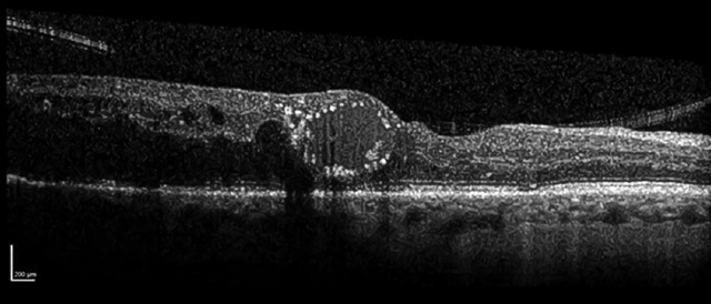
Looking at the OCT image, could you describe the finding in this patient with diabetic macular edema?
ANSWER:
Thanks to everyone who showed interest in the section of question of the month and answered the question. In this month’s question, description of the finding on the OCT image in the patient with diabetic macular edema was asked.
The answer to the question is ‘’ Pearl Necklace Sign’’. The result of the lottery among those who answered the question correctly, the winner of this month’s book prize is Dmytro Martynov. Congratulations to him.
Hyperreflective dots arranged as a contiguous ring along the inner wall of cystoid spaces on the macular OCT scan, termed as the pearl necklace sign, are commonly seen in DMO patients who require intravitreal treatment. With a resolution of edema, hard exudates frequently appear in the same location on the retina, implying that the pearl necklace sign is a precursor to hard exudates, in the majority of cases.
Kshirasagar A, Fiona M, Bipin G, Ajay B. Pearl necklace sign in diabetic macular edema, evaluation and significance. Indian Journal of Ophthalmology 2016;64(11):829-834
https://journals.lww.com/ijo/Fulltext/2016/64110/Pearl_necklace_sign_in_diabetic_macular_edema_.8.aspx

Dymtro MARTYNOV, MD
Medical Center ‘TopMedical’ Zaporizhia, Ukraine
Dr. Dymtro Martynov is graduated from Zaporizhia State Medical University in 2014. After completing his residency in Ophthalmology between 2014-2016 at the Zaporizhia State Medical University, he started to work as an ophthalmologist in the municipal glaucoma office between 2016-2017. He is member of European Glaucoma Society, World Glaucoma Association, EURETINA and ESCRS. He is interested in the retina and glaucoma. He has 25 scientific publications.