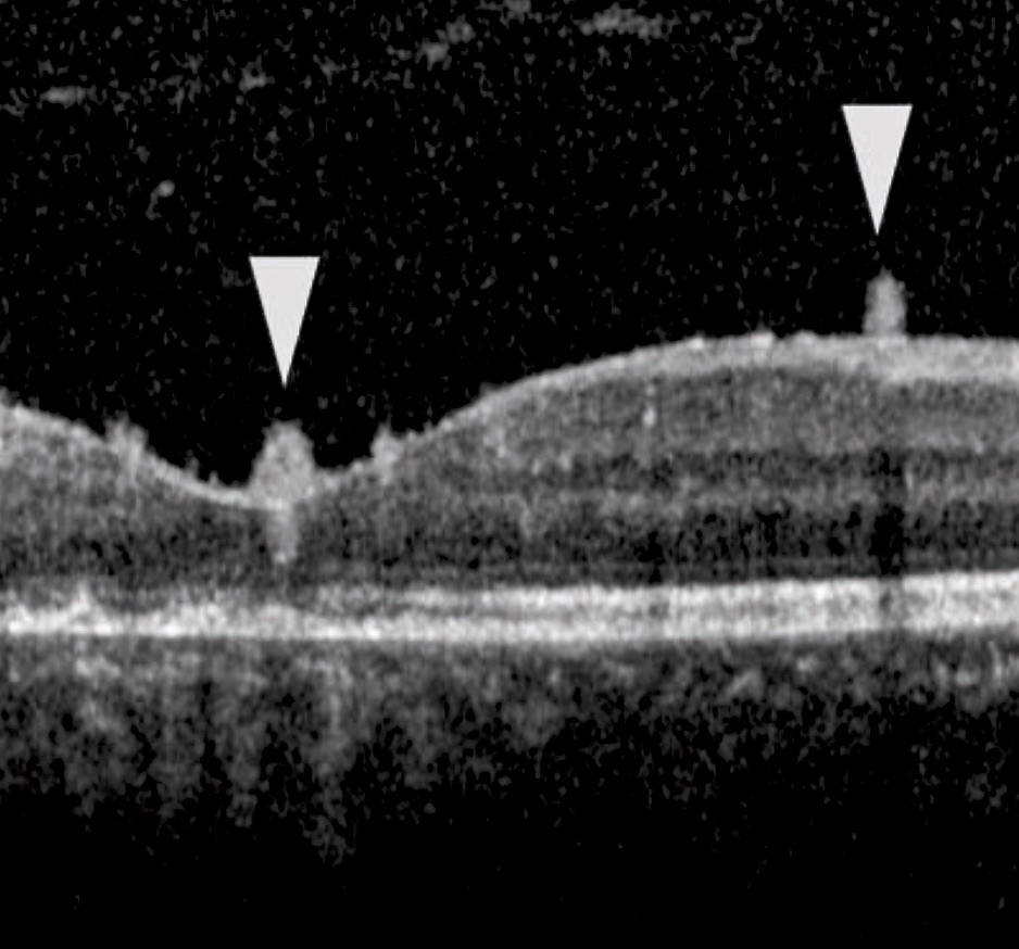
The OCT image of a patient who developed posterior uveitis is shown. Could you determine the diagnosis and describe the OCT findings indicated by arrows of this patient?
Thanks to everyone who showed interest in the section of question of the month and answered the question. In this month’s question, determine the diagnosis and describe the OCT finding in a patient with posterior uveitis were asked.
The answer to the question is ‘’ Hyper-reflective oval deposits within the vitreo-retinal interface in a patient with the active phase of toxoplasmic retino- choroiditis’’. The result of the lottery among those who answered the question correctly, the winner of this month’s book prize is Tuna Ozan, MD. Congratulations to her.
Some diseases tend to develop unique vitritis patterns for which the OCT findings have been characterized. Hyper-reflective oval deposits within the vitreo-retinal interface or along the detached posterior hyaloid have been reported during the active phase of toxoplasmic retino- choroiditis . These deposits migrated within the retina and gradually disappeared as the infection was treated.
https://pubmed.ncbi.nlm.nih.gov/30719788/

Tuna Ozan, MD
Beyoğlu Eye Training and Research Hospital, Istanbul
Dr. Tuna Ozan is graduated from Istanbul University Cerrahpaşa Faculty of Medicine in 2017. She is currently continuing her residency in Ophthalmology at the Beyoglu Eye Training and Research Hospital.