QUESTION:
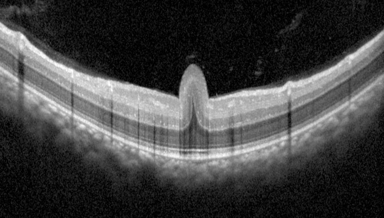
An 8-year-old boy with high hypermetropia presented with BCVA 20/50 OU. Looking at the OCT image, could you describe the finding, and determine the diagnosis?
ANSWER:
Thanks to everyone who showed interest in the section of question of the month and answered the question. In this month’s question, an 8-year- old boy with 20/50 vision with high hypermetropia was presented. Description of the finding on the OCT image and determination of diagnosis were asked.
The answer to the question is “Posterior Microphthalmus/ Macular Fold”. The result of the lottery among those who answered the question correctly, the winner of this month’s book prize is Busenur Gönen, MD. Congratulations to her.
Despite normal anterior segment, posterior microphthalmos is characterized with high hyperopia, shorter axial length and papillomacular retinal fold. Park SH, Ahn YJ, Shin SY, Lee YC. Clinical features of posterior microphthalmos associated with
papillomacular fold and high hyperopia. Clin Exp Optom. 2016;99(6):590-593
https://pubmed.ncbi.nlm.nih.gov/27161391/

Busenur Gönen, MD
Zonguldak Atatürk State Hospital
Dr. Busenur Gönen is graduated from Istanbul University Faculty of Medicine in 2015. After completing his residency in Ophthalmology between 2016-2020 at the Istanbul University Cerrahpasa Faculty of Medicine, she started to work at the Zonguldak Atatürk State Hospital in 2021. She is interested in the retina and ocular oncology. ‘Investigation of Retinal Vascular Changes during Pregnancy Using Optical Coherence Tomography Angiography’, ‘Evaluation of the reasons for the microvascular changes in patients with Fabry disease using optic coherence tomography angiography’ and ‘Investigation of clinical profile of Behçet’s syndrome-related versus idiopathic branch retinal vein occlusion’ are her main articles.