Thanks to everyone who showed interest in the section of question of the month and answered the question. In this month’s question, determine the diagnosis by looking OCT, FAF and IR images in a 52-year-old male patient was asked.
The answer to the question is ‘Macular Telengiectasia Type -2 and Central Serous Chorioretinopathy’.
Despite many ophthalmologists answer the question, unlike the previous months, we did not get a correct answer. In this case, the decrease in vision that developed 1 month ago is due to central serous chorioretinopathy in a patient with diagnosis of macular telangiectasia type-2.
Macular telangiectasia type 2 (MacTel 2) is a bilateral disease characterized by neurodegenerative and vascular changes around the fovea. Degeneration of Müller cells is believed to be the underlying cause. In these eyes, ellipsoid zone disruption initially occurs in the temporal parafoveal area and progresses over time.
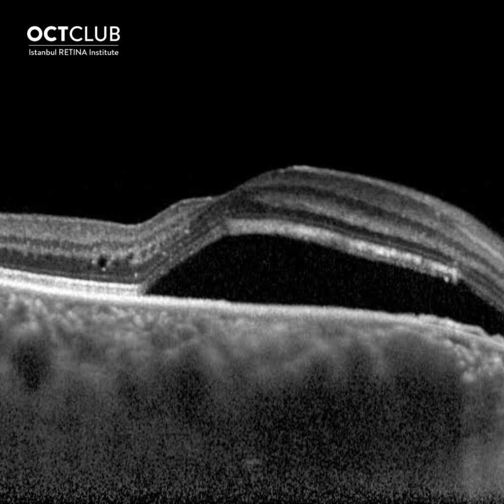
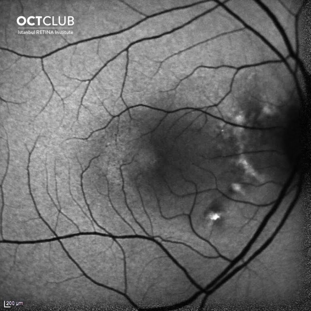
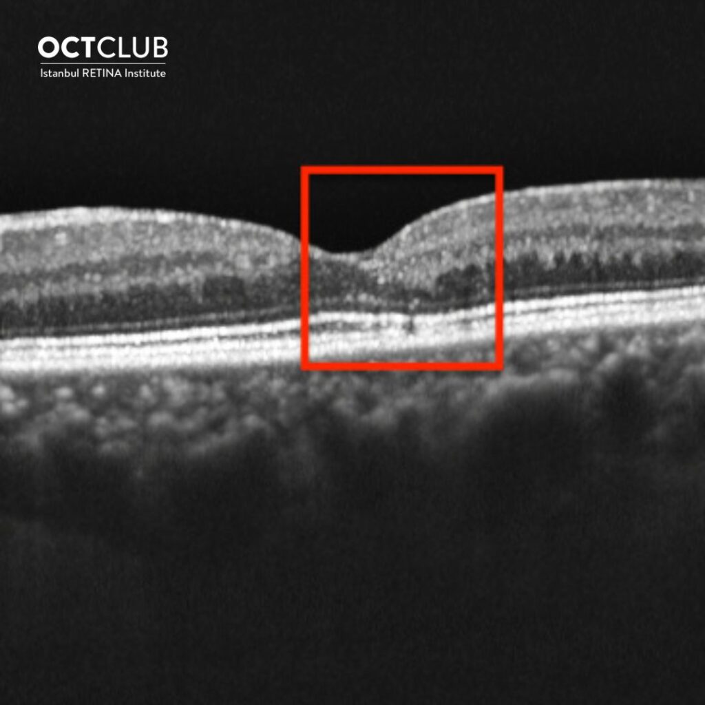
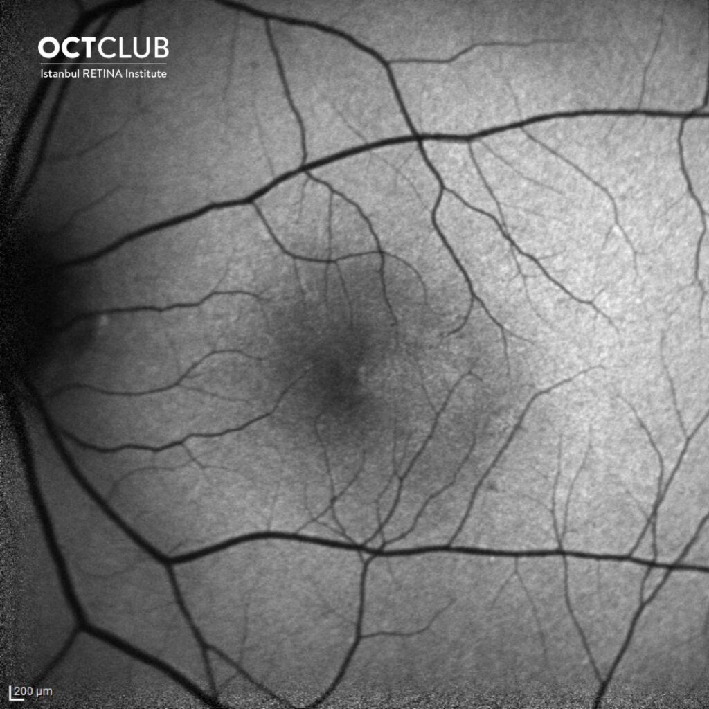
OCT and FAF images show characteristic findings for both MacTel 2 and CSR. In this case, characteristic findings for MacTel 2 are increased autofluorescence and right-angled vessels temporally to the fovea on the FAF image, hyporeflective spaces in the inner retina temporally to the fovea in the right eye, and outer retinal damage in the left eye on the OCT images. Increased choroidal thickness, sensory retinal detachment, and peripapillary shallow irregular pigment epithelial detachment are typical findings for CSR.