Thanks to everyone who showed interest in the section of question of the month and answered the question. In this month’s question, determine the diagnosis by looking OCT, IR images in patient with diagnosis of amd was asked.
The answer to the question is ‘RPE Tear’. The result of the lottery among those who answered the question correctly, the winner of this month’s book prize is Adriana de la Hoz Polo, MD. Congratulations to her.
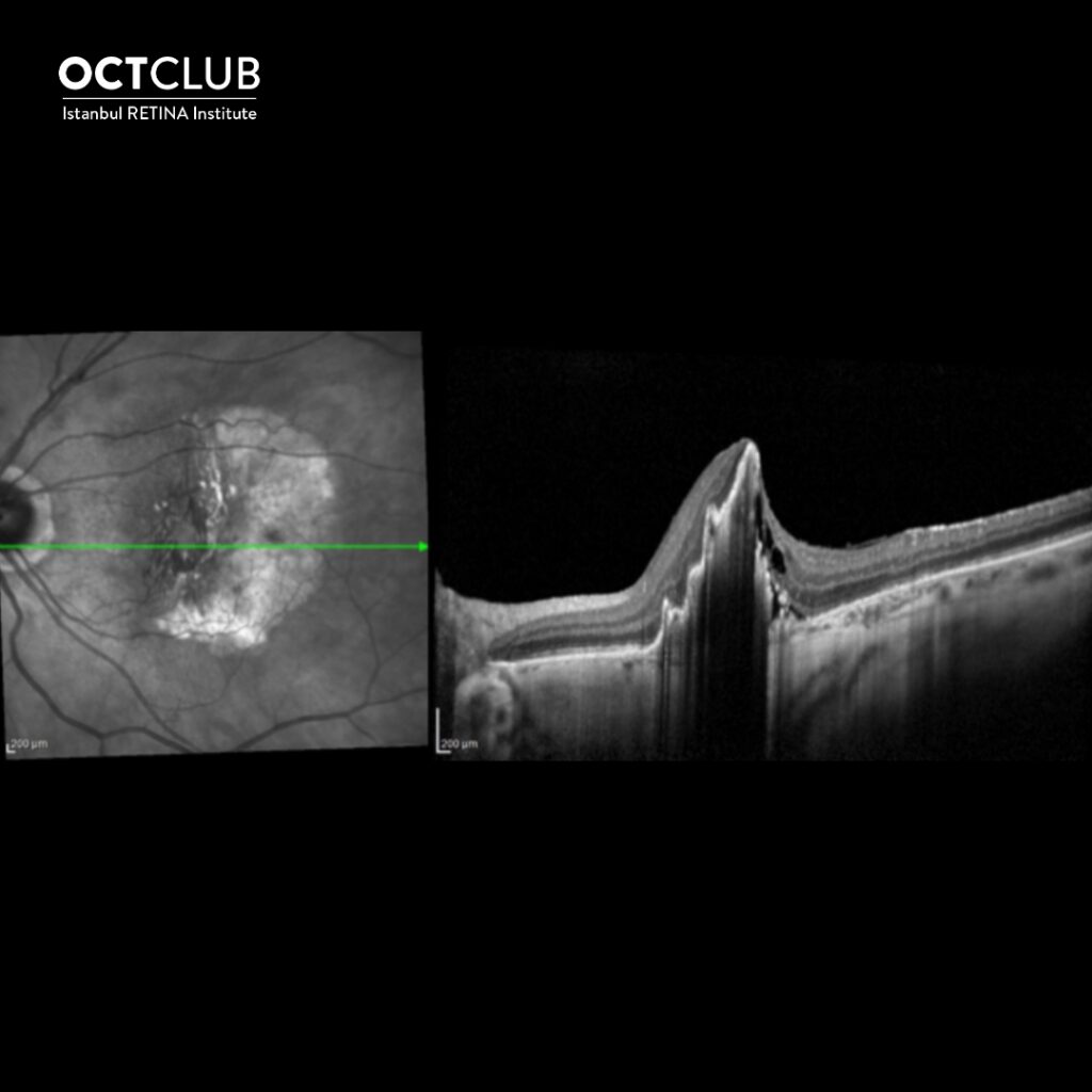
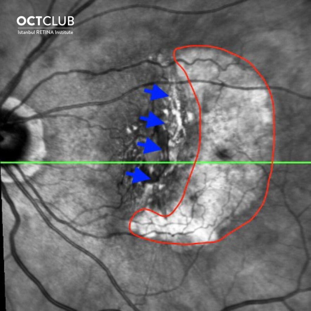
Various eye conditions cause tears in the retinal pigment epithelium (RPE). The most common cause of a RPE tear is vascularized retinal pigment epithelial detachment (PED) in patients with exudative age-related macular degeneration. Although RPE tears can develop spontaneously in vascularized PEDs, most recent cases have been associated with anti-vascular endothelial growth factor (VEGF) injections. The subretinal fluid within the PED applies hydrostatic pressure to the RPE and stretches it. The PED enlarges as the hydrostatic pressure increases. Contraction of the choroidal neovascular membrane adds tractional forces to the RPE monolayer. Especially in larger PEDs, the risk of a RPE tear increases after anti-VEGF therapy owing to increasing contraction of the choroidal neovascular membrane.
https://pubmed.ncbi.nlm.nih.gov/28336128/

Adriana de la Hoz Polo, MD
Universidad CEU San Pablo
Dr. Adriana de la Hoz Polo is graduated from Universidad Autónoma de Madrid in 2015. She is currently working as medical and surgical retinal specialist at the Clínica Miranza, Hospital Universitario de Torrejón and Universidad CEU San Pablo.