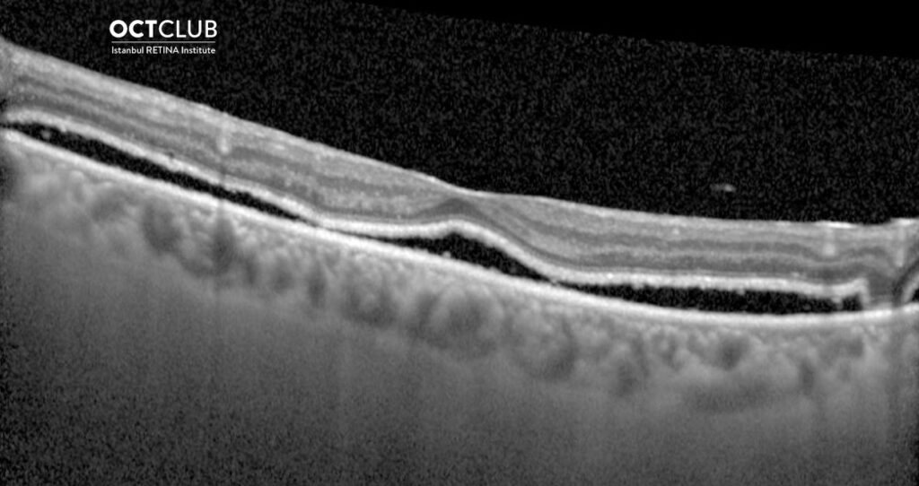
Looking at the OCT image, could you tell the diagnosis in this patient with a history of successful retinal detachment surgery 3 months ago?
Thanks to everyone who showed interest in the section of question of the month and answered the question. In this month’s question, tell the diagnosis by looking at the OCT image in the patient with a history of successful retinal detachment surgery was asked.
The answer to the question is ‘’ Persistent subretinal fluid following successful retinal detachment surgery’’. The result of the lottery among those who answered the question correctly, the winner of this month’s book prize is Merve Şimşek, MD. Congratulations to her/him.
Persistent subfoveolar fluid as seen on optical coherence tomography is a recognized complication of otherwise anatomically successful surgery for macula involving retinal detachments, with reported prevalence between 0 and 94%. Most cases of PSF resolve slowly without adverse sequelae but in a small proportion of cases it is associated with progressive foveal photoreceptor atrophy and loss of visual acuity.
Tee JJL, Veckeneer M, Laidlaw DA. Persistent subfoveolar fluid following retinal detachment surgery: an SD-OCT guided study on the incidence, aetiological associations, and natural history. Eye 2016.
https://www.nature.com/articles/eye2015270

Merve Şimşek,MD
Afyonkarahisar Health Science University Hospital
Dr. Merve Şimşek is graduated fromEskişehir Osmangazi University Faculty of Medicine in 2015. She is currently countinuing her residency in Ophthalmology at the Afyonkarahisar Health Science University Hospital.