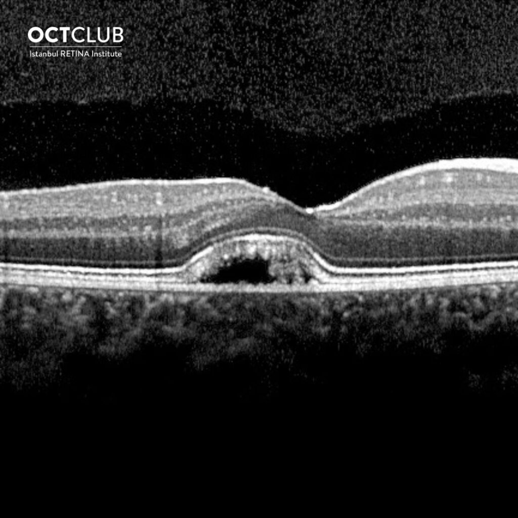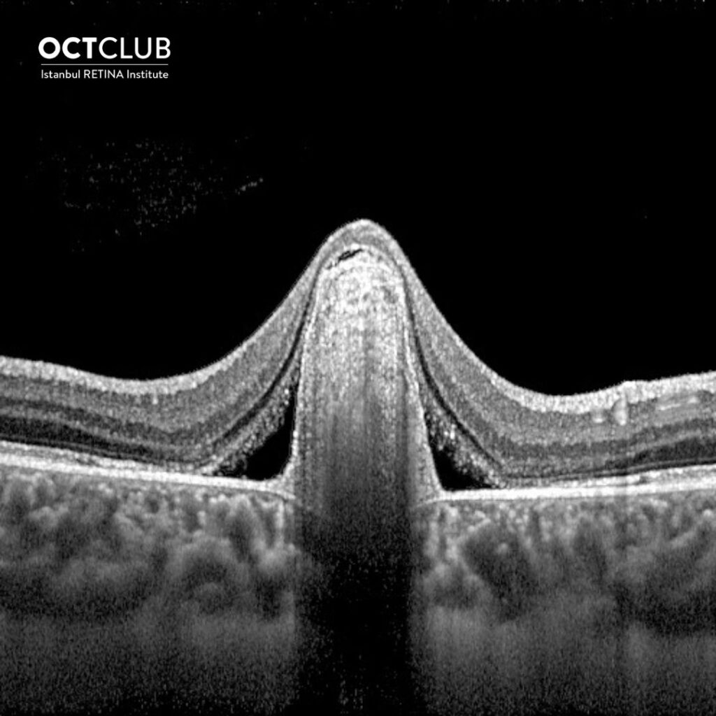

Looking at the optical coherence tomography images, could you tell the diagnosis in this young male patient?
Thanks to everyone who showed interest in the section of question of the month and answered the question. In this month’s question, tell the diagnosis by looking at the OCT image was asked.
The answer to the question is ‘’ Best Disease’’. The result of the lottery among those who answered the question correctly, the winner of this month’s book prize is Joao Pedro Teixeira Marques,MD,MSc,FEBO. Congratulations to him.
Although the appearance of lipofuscin accumulation between the retinal pigment epithelium and the outer retina is characteristic of Best Disease, presence of hyperreflective material between the RPE and Bruch’s membrane, with serous macular detachment, may be the first presenting finding in some patients. This hyperreflective RPE elevation on SD-OCT may represent fibrosis, quiescent neovascularization, or active neovascularization, and multimodal imaging techniques are very important for differentiation of these lesions.
Sayman Muslubas I, Arf S, Hocaoglu M, Ersoz MG, Karacorlu M. Best Disease Presenting as Subretinal Pigment Epithelium Hyperreflective Lesion on Spectral-Domain Optical Coherence Tomography: Multimodal Imaging Features. Eur J Ophtalmol 2021 Online ahead of print
https://pubmed.ncbi.nlm.nih.gov/34806463/

Joao Pedro Teixeira Marques, MD, FEBO, MSc
Centro Hospitalar e Universitario de Coimbra (CHUC), Coimbra, Portugal
Dr. Joao Pedro Teixeira Marques is graduated from University of Coimbra in 2010. He completed his residency in Ophthalmology between 2012-2015 at the Centro Hospitalar e Universitario de Coimbra (CHUC). He is ophthalmology specialist and investigator at the Retina Department of Centro Hospitalar e Universitario de Coimbra, and head of the Retinal Dystrophies Clinic at CHUC. He is member of the EVICR.net Retinal Dystrophies Expert Committee. He has 57 scientific publications.


