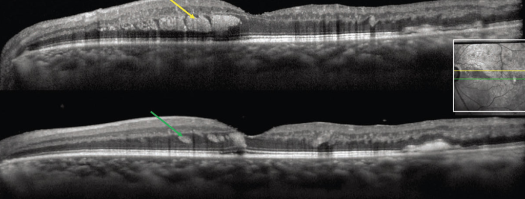
Thanks to everyone who showed interest in the section of question of the month and answered the question. In this month’s question, identify the OCT finding in patient with branch retinal vein occlusion was desired.
The answer to the question is ‘Henle Fiber Layer Hemorrhage. The result of the lottery among those who answered the question correctly, the winner of this month’s book prize is Maria Vittoria Cicinelli, MD. Congratulations to her.
OCT findings of blood located within Henle fiber layer, referred to as Henle fiber layer hemorrhage or HH, have been described as characteristic hyperreflectivity from the hemorrhage delineated by the obliquely oriented fibers in the Henle layer. Henle hemorrhage may result from a wide variety of pathologies and can be classified as secondary to local vascular abnormalities of the deep capillary plexus, choroidal vascular abnormalities or disorders affecting central venous pressure.
https://www.retina-specialist.com/article/can-you-recognize-these-novel-oct-signs

Maria Vittoria Cicinelli, MD
Vita-Salute San Raffaele University, Milano
Dr Maria Vittoria Cicinelli is graduated from Vita-Salute San Raffaele University Faculty of Medicine in 2013. After completing her residency in Ophthalmology between 2014-2019 at the Vita-Salute San Raffaele University, she started to work at same hospital in 2020, as an ophtalmologist. She is interested in medical retina and uveal diseases.


